Visual reference
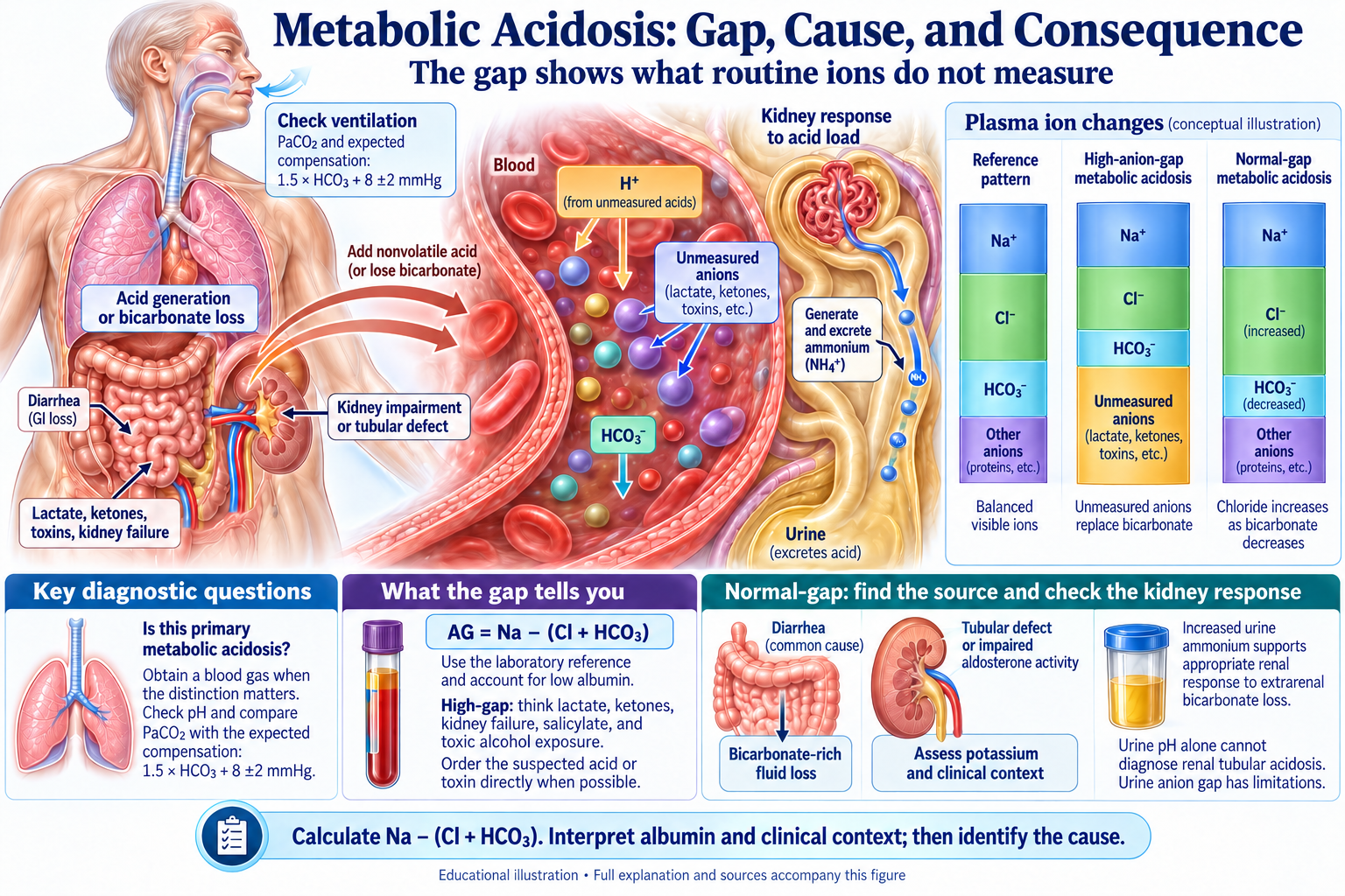
Browse the visual reference library · Download lesson Markdown
Treat urgent threats while you evaluate
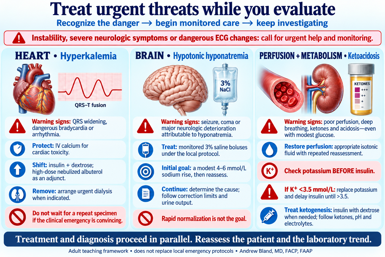
Recognize danger, begin monitored care and keep investigating in parallel.
Lactated Ringer’s versus normal saline
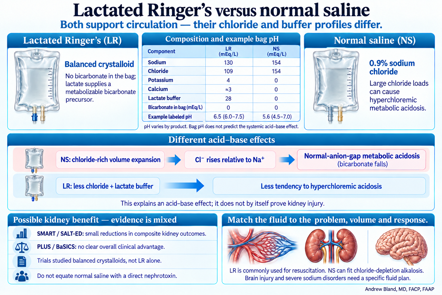
Different chloride and buffer profiles; possible kidney benefit with mixed clinical evidence.
Composition helps explain the acid–base effect
Typical LR contains sodium 130, chloride 109, potassium 4, calcium approximately 3 and lactate 28 mEq/L; formulation details vary. Normal saline contains sodium and chloride 154 mEq/L each, with no potassium, calcium or buffer. LR is commonly grouped with isotonic resuscitation crystalloids, although its measured osmolarity is slightly below plasma.
Neither bag contains bicarbonate
Both LR and NS contain zero bicarbonate in the bag. LR provides lactate as a metabolizable bicarbonate precursor; NS has no buffer anion. Saline-associated hyperchloremic acidosis is sometimes described as dilutional or expansion acidosis, but chloride-rich composition—not volume expansion alone and not the acidity of the bag—is the central teaching point.
Example bag pH is not the systemic acid–base effect
Example manufacturer labels list LR at pH 6.5 (range 6.0–7.5) and NS at pH 5.6 (4.5–7.0). Values vary by product and container. Bag pH is unitless and does not measure the delivered titratable acid load or predict the patient’s response; the chloride burden, metabolizable anions, dose and patient physiology matter far more.
Why chloride rises while bicarbonate falls
A large chloride-rich saline load can raise chloride relative to sodium, lower the plasma strong-ion difference and produce a normal-anion-gap metabolic acidosis. This is not a claim that saline directly triggers bicarbonate loss at a particular tubular transporter. LR supplies less chloride and lactate that can be metabolized to a bicarbonate equivalent; the lactate anion is not an infusion of lactic acid.
What the trials support—and what they do not
SMART found a small reduction in a composite of death, new dialysis or persistent renal dysfunction with balanced crystalloids. SALT-ED found a similar secondary kidney-composite signal, while its primary hospital-free-days outcome was unchanged. The large blinded PLUS and BaSICS trials did not establish a clear overall advantage in their primary mortality outcomes; PLUS also found similar kidney outcomes. These trials tested balanced-crystalloid strategies, sometimes LR or Plasma-Lyte and sometimes Plasma-Lyte alone. They do not prove that LR universally prevents AKI or that normal saline is a direct nephrotoxin.
Choose the fluid for the clinical problem
A possible modest kidney benefit and less hyperchloremic acidosis support considering balanced crystalloids when appropriate. Chloride-depletion alkalosis may favor saline. Brain injury, severe sodium disorders, compatibility issues and specific electrolyte problems require a tailored plan. Fluid volume, indication and repeated reassessment remain central; these resuscitation studies should not be treated as proof of the best maintenance regimen.
Supporting evidence
🚨 Emergency Management Priority
Critical pH Thresholds
- pH < 7.1: Life-threatening - immediate intervention
- pH < 7.2: Severe - aggressive treatment
- pH 7.2-7.35: Moderate - treat underlying cause
- Ensure adequate ventilation (respiratory compensation)
- IV access and volume status assessment
- Calculate anion gap and assess for mixed disorders
- Treat underlying cause (DKA, lactic acidosis, toxins)
- Consider bicarbonate ONLY if pH <7.15 with shock
🔬 The ABC Method: Systematic Acid-Base Analysis
Step A: Assess pH
- Acidemia: pH < 7.40
- Alkalemia: pH > 7.40
- Remember: Acidosis ≠ Acidemia
- Note: Can have acidosis with normal/high pH due to compensation
Step B: Primary Disorder
- Metabolic: Primary HCO₃⁻ change
- Respiratory: Primary pCO₂ change
- Mixed: Both systems affected
- Key: Which change explains the pH?
Step C: Check Compensation
- Never complete: pH always abnormal
- Never overcompensates: pH doesn't cross 7.40
- Winter's Formula: Expected pCO₂ = 1.5 × [HCO₃⁻] + 8 ± 2
- If overcompensation: Mixed disorder present
🧮 Anion Gap: The Diagnostic Key
Anion Gap Calculation
Normal AG: 8-12 mEq/L (modern analyzers)
Traditional AG: 12±4 mEq/L (older labs)
⚠️ Know your lab's normal range!
High Anion Gap Acidosis (MUDPILES)
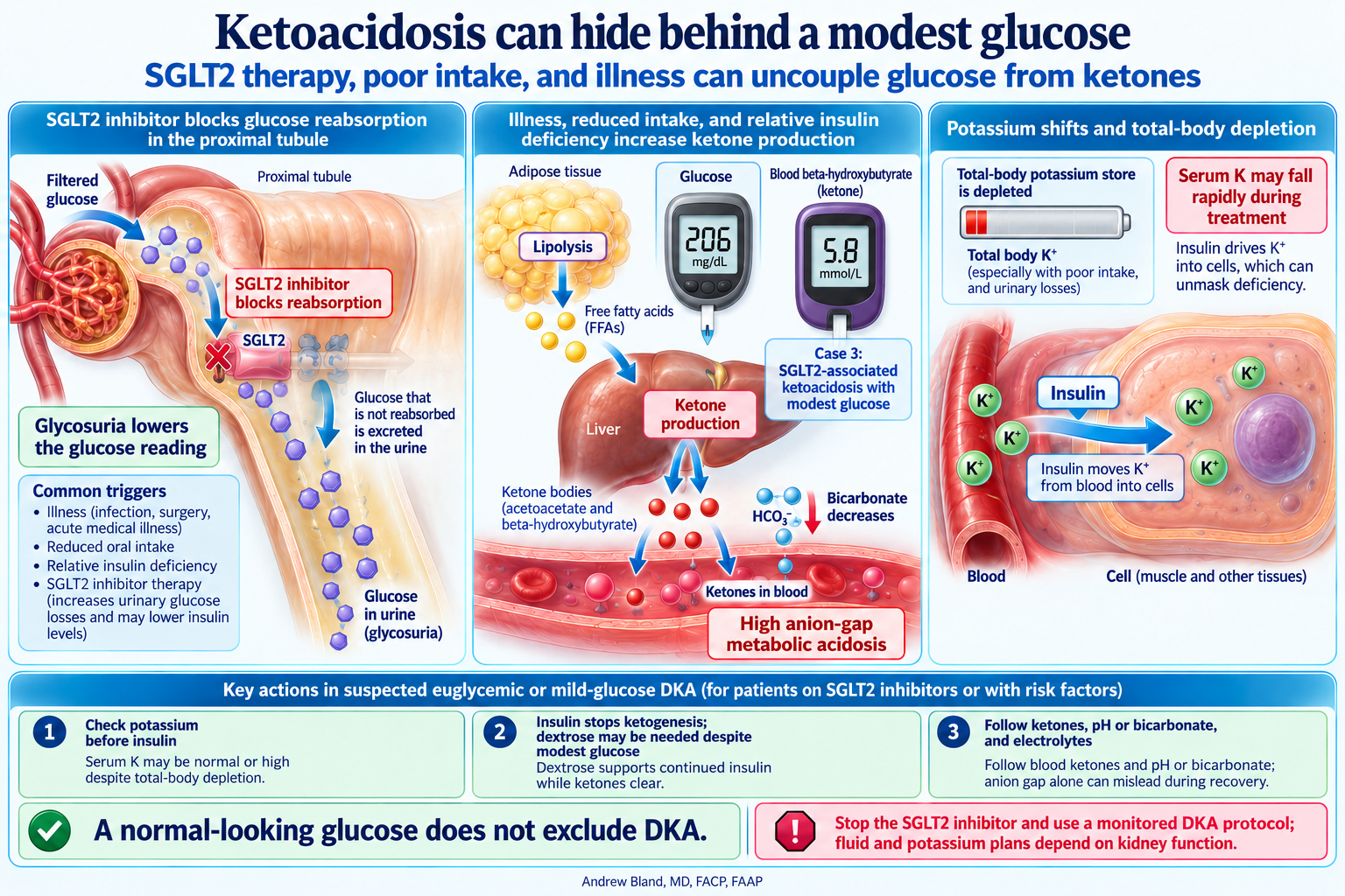
SGLT2 therapy, poor intake, and illness can uncouple glucose from ketones
- M: Methanol
- U: Uremia (BUN >100)
- D: DKA, alcoholic ketoacidosis
- P: Propylene glycol (IV Ativan!)
- I: Isoniazid, Iron
- L: Lactic acidosis (most common)
- E: Ethylene glycol
- S: Salicylates
Normal AG Acidosis (USED CARP)
Normal Kidney Function (UAG < 0):
- U: Ureterosigmoidostomy
- S: Small bowel fistulae
- E: Extra Cl⁻ (normal saline)
- D: Diarrhea (most common)
Impaired Kidney Function (UAG > 0):
- C: Chronic kidney disease
- A: Acetazolamide, Addison's
- R: Renal tubular acidosis
- P: Protein overfeeding
Urinary Anion Gap (UAG) for Normal AG Acidosis
UAG < 0 (Negative)
Meaning: Appropriate NH₄⁺ production
Cause: Extra-renal HCO₃⁻ loss
Examples: Diarrhea, fistulae, saline administration
UAG > 0 (Positive)
Meaning: Impaired NH₄⁺ production
Cause: Renal acid excretion defect
Examples: RTA, CKD, hypoaldosteronism
🔄 Delta-Delta Ratio: Detecting Mixed Disorders
The Delta-Delta Calculation
Δ AG = Measured AG - Normal AG (10)
Δ HCO₃⁻ = Normal HCO₃⁻ (24) - Measured HCO₃⁻
Delta/Delta Ratio = Δ AG / Δ HCO₃⁻
Ratio < 1.0
Interpretation: Mixed disorder
- High AG metabolic acidosis
- PLUS normal AG metabolic acidosis
Ratio 1.0-2.0
Interpretation: Pure disorder
- High AG metabolic acidosis only
- Expected compensation pattern
Ratio > 2.0
Interpretation: Mixed disorder
- High AG metabolic acidosis
- PLUS metabolic alkalosis
Alternative: Potential Bicarbonate Method
Concurrent NAG acidosis
Pure AG acidosis
Concurrent alkalosis
🧮 Interactive Acid-Base Calculator
📋 Clinical Case Applications
Case 1: ICU Patient with Sepsis
Clinical: 55M, severe sepsis, received 15L normal saline, on lorazepam drip
Labs: pH 7.10, pCO₂ 19, HCO₃⁻ 6, Na 135, Cl 110, UAG -55
Analysis:
- Severe acidemia (pH 7.10)
- Primary metabolic acidosis (HCO₃⁻ 6)
- Appropriate respiratory compensation (expected pCO₂ 17±2)
- AG = 135-110-6 = 19 (elevated)
- Delta/Delta = 9/18 = 0.5 (<1 = mixed)
Diagnosis: AG acidosis (propylene glycol from lorazepam) + NAG acidosis (NS-induced)
Case 2: DKA with Fungal Sepsis
Clinical: 60M, DM, fungal sepsis
Labs: pH 7.01, pCO₂ 39, HCO₃⁻ 10, glucose 480, β-hydroxybutyrate 3, lactate 8
Analysis:
- Severe acidemia with inadequate respiratory compensation
- Expected pCO₂ = 1.5×10 + 8 = 23 ± 2 (actual 39 = respiratory acidosis too)
- Multiple causes: DKA + lactic acidosis + respiratory failure
- Triple acid-base disorder
Management: Insulin, fluids, ventilatory support, treat sepsis
Case 3: C. diff Colitis vs RTA
Clinical: 67F, C. diff colitis
Labs: pH 6.91, pCO₂ 40, HCO₃⁻ 8, Na 135, Cl 110, UAG -40
Analysis:
- Severe acidemia with minimal respiratory compensation (concerning)
- AG = 17 (elevated, likely from uremia/dehydration)
- UAG = -40 (negative = appropriate NH₄⁺ production)
- Negative UAG confirms diarrheal losses
Diagnosis: Mixed uremic acidosis + diarrhea-induced NAG acidosis
💊 Treatment Principles
Primary Rule: Treat the Underlying Cause
Correcting pH without addressing the underlying pathophysiology is ineffective and potentially harmful.
High AG Metabolic Acidosis
- DKA: Insulin, fluids, electrolyte replacement
- Lactic acidosis: Improve tissue perfusion
- Uremia: Dialysis for severe cases
- Toxic ingestions: Specific antidotes, dialysis
- Ketoacidosis: Thiamine, glucose, insulin
Normal AG Metabolic Acidosis
- Diarrhea: Volume and electrolyte replacement
- RTA: Alkali therapy (K-citrate preferred)
- Saline-induced: Switch to balanced crystalloids
- Hypoaldosteronism: Mineralocorticoid replacement
⚠️ Bicarbonate Therapy: Use Sparingly
Consider Only When:
- pH < 7.15 with hemodynamic instability
- Severe hyperkalemia with ECG changes
- Certain toxic ingestions (salicylates, methanol, TCA)
- Severe RTA with failure to thrive
Risks of Bicarbonate Therapy:
- Paradoxical CSF acidosis
- Hypokalemia and hypocalcemia
- Volume overload
- Rebound alkalosis
- Shift of O₂-hemoglobin curve
HCO₃⁻ deficit = 0.5 × weight (kg) × (desired HCO₃⁻ - current HCO₃⁻)
Give 1/2 to 1/3 of calculated deficit over 3-4 hours
🎯 Key Learning Points
Systematic Approach
- Always use the ABC method for acid-base analysis
- Compensation is never complete or overcompensating
- Check for mixed disorders in all complex cases
- Use Winter's formula to assess appropriate compensation
Anion Gap Mastery
- Know your lab's normal AG range (8-12 vs 12±4)
- Always correct for albumin in critically ill patients
- Use UAG to differentiate renal vs GI causes of NAG acidosis
- Delta-delta ratio reveals mixed metabolic disorders
Treatment Priority
- Always treat the underlying cause, not just the pH
- Bicarbonate therapy is rarely indicated and often harmful
- Support ventilation for respiratory compensation
- Address volume status and electrolyte abnormalities