⚡ Urgent Evaluation Protocol
1
Acute Kidney Failure: Rapid decline + bilateral masses → Urgent biopsy, chemotherapy consultation
2
B-symptoms: Fever, night sweats, weight loss → Hematologic evaluation
3
Hypercalcemia: Malignancy-associated → Bisphosphonates, fluid resuscitation
4
Multiple Renal Masses: Known primary malignancy → Metastatic workup
🎯 Renal Lymphoma & Metastases: Key Facts
📊 Renal Lymphoma
5% CT detection, 30-60% autopsy involvement - Often clinically silent
🫁 Primary Origins
Lung, breast, melanoma, colon cancers most commonly metastasize to kidney
🔬 B-cell NHL
Most common hematologic malignancy involving the kidney
🏥 Primary Rare
Primary renal lymphoma <70 cases reported in literature
🩺 Renal Lymphoma
- Incidence: 5% of lymphoma patients by CT evaluation
- Autopsy Studies: 30-60% involvement found post-mortem
- Detection Gap: Often clinically silent, imaging underdetects
- Primary Form: Rare, <70 cases reported in literature
- Secondary: More common, indicates advanced disease
- Histology: B-cell non-Hodgkin lymphoma most common
🖼️ Imaging Patterns
- Bilateral Enlargement: Most common presentation
- Hypoenhancing Masses: Poor contrast enhancement typical
- Multiple Nodules: Infiltrative disease pattern
- Retroperitoneal LAD: Associated lymphadenopathy
- FDG Avidity: Intense uptake on PET imaging
- Preservation of Shape: Reniform shape often maintained
🎯 Metastatic Disease to the Kidney
Primary Tumor Origins
- Lung Cancer: Most common source of renal metastases
- Melanoma: High propensity for kidney involvement
- Breast Cancer: Common in advanced disease
- Colon Cancer: Late manifestation of disease
- Thyroid Cancer: Rare but reported
- Gastric Cancer: Occasional kidney involvement
Clinical Considerations
- Autopsy Frequency: Common finding post-mortem
- Clinical Presentation: Usually asymptomatic
- Atypical Imaging: Suspicious for metastases
- Constitutional Symptoms: Weight loss, fatigue
- Advanced Disease: Indicates poor prognosis
- Systemic Treatment: Focus on primary malignancy
🔍 Diagnostic Evaluation Protocol
1
Clinical History: Prior malignancy, B-symptoms, constitutional symptoms
2
Imaging Evaluation: CT chest/abdomen/pelvis, PET if lymphoma suspected
3
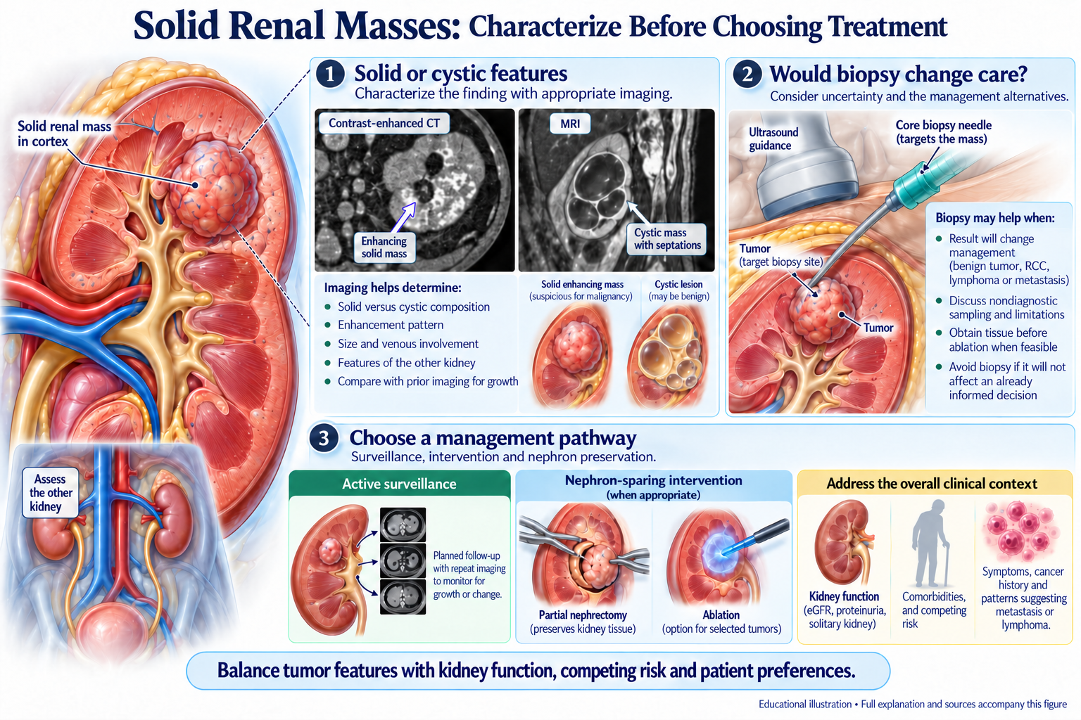
Tissue Diagnosis: Image-guided biopsy essential for confirmation
4
Laboratory Studies: LDH, β2-microglobulin, flow cytometry
5
Staging Workup: Bone marrow biopsy if hematologic malignancy
🧮 Renal Mass Differential Diagnosis Tool
Most Likely Diagnosis
Primary Renal Cell Carcinoma
Recommend tissue biopsy for definitive diagnosis
💊 Lymphoma Treatment
- Systemic Chemotherapy: Primary treatment modality
- No Surgery: Surgical resection not indicated
- Response Rates: Good response to appropriate chemotherapy
- Renal Function: Complete recovery uncommon
- Advanced Disease: Kidney involvement indicates poor prognosis
- Supportive Care: Manage CKD complications
🎯 Metastatic Disease Management
- Primary Focus: Treat underlying primary malignancy
- Systemic Therapy: Chemotherapy based on primary tumor
- Local Treatment: Usually not indicated
- Palliative Care: Symptom management priority
- Prognosis: Generally poor when kidney involved
- Supportive Measures: CKD management, electrolyte balance
🔬 Renal Mass Biopsy Decision Framework
✅ Strong Indications for Biopsy
- Prior history of lymphoma or other malignancy
- Bilateral or multiple renal masses
- Atypical imaging characteristics
- Constitutional symptoms present
- Associated lymphadenopathy
- Acute kidney injury with mass
⚠️ Biopsy Considerations
- Image-guided core needle biopsy preferred
- Adequate tissue for flow cytometry
- Avoid fine needle aspiration
- Consider bleeding risk assessment
- Multidisciplinary team discussion
- Patient counseling about alternatives
🎯 Key Learning Points
High Index of Suspicion
Consider lymphoma or metastases in patients with atypical renal masses, especially with known malignancy history.
Tissue Diagnosis Essential
Image-guided biopsy is crucial for accurate diagnosis and appropriate treatment planning.
Systemic Disease
Renal involvement usually indicates advanced systemic disease requiring oncologic management.
Multidisciplinary Care
Optimal outcomes require coordination between nephrology, oncology, and hematology specialists.