Why This Review Exists
Two drugs reached the US market within eight months of each other. VOYXACT (sibeprenlimab-szsi) — a humanized IgG2 anti-APRIL monoclonal — received FDA accelerated approval on 25 November 2025. TRUTAKNA (atacicept-vymj) — a TACI-Fc fusion protein neutralizing both BAFF and APRIL — followed on 7 July 2026, the first dual-target agent approved for this disease.
Both are promoted as acting "furthest upstream." Commercial materials have begun describing the BAFF/APRIL axis as sitting before Hit 1 of the four-hit model. That framing deserves scrutiny, because it makes a strong implicit promise: block this node, and the pathogenic cascade never starts.
The biology is more interesting than the slogan, and considerably less tidy.
The pathogenic glycoform is galactose-deficient IgA1 (Gd-IgA1) — the hinge-region O-glycans lack terminal galactose, exposing N-acetylgalactosamine. It is not mannose-deficient. The partner cytokine is BAFF (B-cell activating factor, TNFSF13B); APRIL is TNFSF13.
Section 1 — Is "Pre-Hit 1" a Real Designation?
Short answer: no. It is sponsor positioning, not a peer-reviewed renumbering of the model.
No primary source in the IgAN literature designates a formal "Hit 0" or "pre-Hit 1." The regulatory and commercial language describes BAFF and APRIL as "the two cytokines thought to sit furthest upstream in the disease's pathology" [22]. That is a spatial metaphor, not a taxonomy.
What the peer-reviewed literature actually says is narrower and more specific. Cheung and colleagues, in the definitive review of this axis, place BAFF and APRIL as the key mediators of Hit 1 and Hit 2 — the cytokine inputs that drive both Gd-IgA1 production and the subsequent autoantibody response [1]. They are not a separate antecedent event. They are the signaling machinery inside the first two hits.
This distinction is not pedantic. It has three consequences:
First, APRIL is not a single upstream valve — it feeds two hits at once. Blocking it is not "preventing Hit 1." It is partially attenuating Hits 1 and 2 simultaneously.
Second, there is a genuine tier above APRIL, and no approved agent touches it. Makita and colleagues showed that TLR9 activation drives aberrant IgA glycosylation through APRIL and IL-6, and stated the finding explicitly: "APRIL and IL-6 pathways each independently mediated TLR9-induced overproduction of Gd-IgA1" [2]. Innate mucosal sensing sits above APRIL. IL-6 runs parallel to it. If anything deserves the label "pre-Hit 1," it is TLR9/microbial sensing.
Third, the newest pathogenesis work adds pathways that bypass Gd-IgA1 entirely. Nihei and Suzuki have identified IgA-type anti-mesangial cell antibodies (IgA-MESCA) targeting the mesangial surface antigens β2-spectrin and CBX3 [3]. If glomerular-specific autoantibodies contribute independently, upstream IgA suppression addresses only part of the disease.
"Furthest upstream" is true relative to the endothelin and complement agents. It is false as an absolute claim. TLR9 and IL-6 sit above APRIL, and IL-6 reaches Gd-IgA1 without passing through it. That single fact predicts most of what follows in this review.
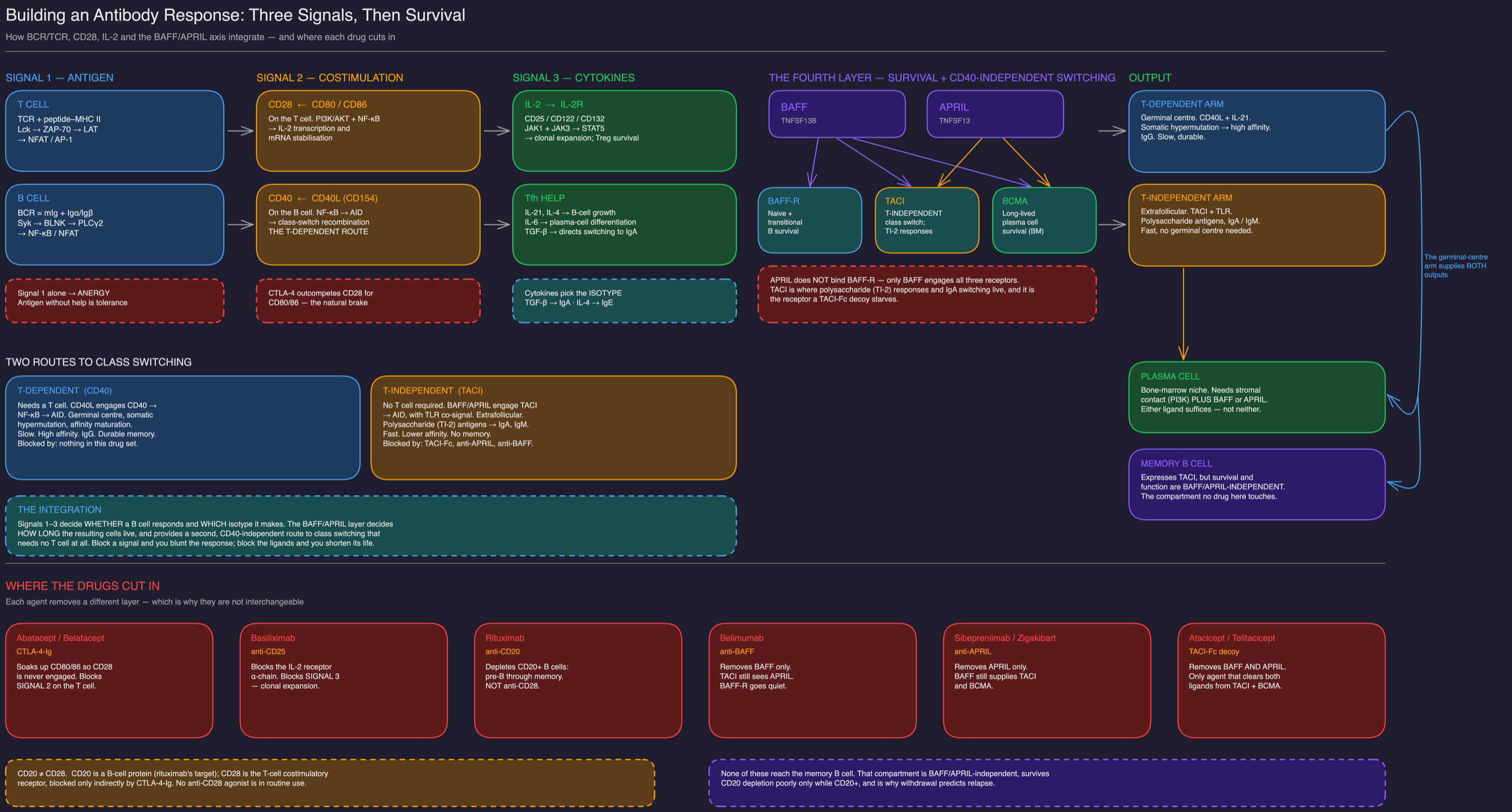
Section 2 — Does APRIL Blockade Preferentially Target Gd-IgA1?
Short answer: no. The selectivity is by isotype, not by glycoform. Nothing in this class reads the hinge-region O-glycan.
This is the most consequential misconception about the drug class, and the preclinical data settle it cleanly.
Dulos and colleagues characterized zigakibart (an anti-APRIL IgG4 monoclonal in phase 3) and reported that it "selectively reduced circulating IgA and IgM to a greater extent than IgG" and "inhibited IgA+ and IgM+ to a greater extent than IgG+ human plasma cells in vitro" [4]. The discrimination is between isotypes — IgA and IgM versus IgG. There is no mechanism by which an anti-APRIL antibody distinguishes a galactosylated IgA1 hinge from a galactose-deficient one.
The clinical pharmacology matches. In the zigakibart phase 1/2 study, healthy volunteers showed "durable reductions in levels of free APRIL, IgA and IgM, and to a lesser extent, IgG," and IgAN patients showed "rapid and durable reductions in IgA, galactose-deficient IgA (Gd-IgA1), and IgM levels, with a modest reduction in IgG" [5]. Gd-IgA1 falls because total IgA falls. It is a passenger, not the target.
The 67% Ceiling
Here the numbers become genuinely instructive. Line up Gd-IgA1 reduction across mechanisms and trials:
| Agent | Mechanism | APRIL suppression | Gd-IgA1 reduction | Source |
|---|---|---|---|---|
| Sibeprenlimab 400 mg | APRIL-only | 95.8% (wk 48) | 67.1% (wk 48) | VISIONARY [6] |
| Atacicept 150 mg | Dual BAFF/APRIL | not reported | 68% (wk 36) | ORIGIN 3 [22] |
| Atacicept 150 mg | Dual BAFF/APRIL | not reported | −66% ± 2% (wk 96) | ORIGIN 2 OLE [8] |
| Telitacicept 240 mg | Dual BAFF/APRIL | not reported | 50.4% (wk 24) | Phase 2 biomarker [9] |
The VISIONARY pairing is the striking one. Sibeprenlimab removed 95.8% of circulating APRIL and achieved 67.1% Gd-IgA1 reduction [6]. Near-total ligand neutralization left roughly a third of the pathogenic glycoform standing.
Dual blockade does not break through it either — atacicept lands at 66–68% across two independent readouts [8,22]. Three agents, two mechanisms, four timepoints, and the answer keeps converging near two-thirds.
The VISIONARY publication closes the argument, and with confidence intervals the label does not carry. At week 48 [6]:
| Analyte | Change from baseline | 95% CI |
|---|---|---|
| APRIL | −95.8% | 93.9 to 97.7 |
| IgM | −74.5% | 73.1 to 75.9 |
| IgA | −68.8% | 67.2 to 70.5 |
| Gd-IgA1 | −67.1% | 62.8 to 71.3 |
| IgG | −35.0% | 32.8 to 37.3 |
The two intervals that matter overlap almost completely: IgA 67.2–70.5 and Gd-IgA1 62.8–71.3. These are not merely similar point estimates — they are statistically inseparable. Nothing is selecting the galactose-deficient hinge; the isotype is being lowered and the glycoform rides down with it. The authors state the same conclusion plainly: "total IgA and IgM levels were reduced by approximately 70% with sibeprenlimab, whereas IgG levels were reduced by 35%" [6].
Note also that the suppression is reversible and dose-dependent. In the first-in-human study, APRIL, IgA, Gd-IgA1, IgG and IgM were all "reversibly suppressed in a dose-dependent manner, with a dose-response in time to recovery" [54].
There is an APRIL-independent Gd-IgA1 floor of roughly one-third, and it is not overcome by adding BAFF blockade. Makita's IL-6 pathway is the most likely explanation [2]. Expect a residual Gd-IgA1 burden in every patient on these drugs — and manage proteinuria accordingly rather than assuming the source is switched off.
Section 3 — Are These Signals Restricted to IgA-Committed B Cells?
Short answer: no. The receptors are pan-B-lineage and stage-dependent. TACI is on T cells as well.
The three receptors are not IgA-specific in any sense.
Zhang and colleagues mapped expression across human tonsillar B-cell differentiation and found BAFF-R dominant on naive B cells, with TACI and BCMA induced progressively as germinal-center B cells differentiate toward plasma cells [10]. The receptor repertoire tracks differentiation stage, not isotype commitment.
APRIL binds BCMA on B cells and TACI on B and T cells [11]. Barone and colleagues found APRIL, TACI, and BCMA distributed through GALT, lamina propria, and the epithelium of stomach, small and large intestine, and rectum [12]. This is general mucosal immune architecture.
So where does the apparent IgA selectivity come from? Three contextual factors, not receptor restriction:
- Co-localization. Barone showed that APRIL and its receptors appear alongside activation-induced cytidine deaminase (AID) — absolutely required for class switching — only in GALT, not in lamina propria [12]. Anatomy concentrates the effect where IgA switching happens.
- Switch-direction bias. APRIL by itself induces IgA and IgG1 switching in CD40-deficient B cells [11] — it is intrinsically an IgA-directing signal in the mucosal context.
- Receptor upregulation in disease. Zhai and colleagues found elevated plasma APRIL in IgAN alongside higher BCMA and TACI expression on patient B lymphocytes, with plasma APRIL correlating with Gd-IgA1 levels and with worse proteinuria and eGFR [13].
Because these receptors are pan-B-lineage, the therapeutic effect cannot be anatomically contained. Loss-of-function TNFRSF13B (TACI) mutations cause common variable immunodeficiency and selective IgA deficiency in humans — B cells from these patients "did not produce IgG and IgA in response to the TACI ligand APRIL" [14]. Pharmacologic TACI-axis blockade partially phenocopies a known human immunodeficiency. See Section 7 — this is not a theoretical concern.
Section 4 — Does It Inhibit Memory B Cells?
Short answer: no — and this is the single most important limitation of the entire class.
Benson and colleagues addressed exactly this question and the finding is unambiguous: "long-lived B(MEM) cell survival and function are completely independent of BAFF or APRIL. Thus, B(MEM) cells represent the only mature B2 lineage subset whose survival is independent of these ligands" [15].
The only B-cell compartment that survives without BAFF or APRIL is the memory compartment. Every other mature B2 subset depends on these ligands. Memory does not.
The clinical implications are direct and underappreciated:
- The Gd-IgA1-committed memory pool persists intact. These drugs suppress output; they do not erase the immunologic program that generates it.
- Relapse on withdrawal should be the default expectation. Memory B cells survive the drug and can re-differentiate once the ligand returns.
- These are chronic suppressive therapies, not curative or remittive ones. Nothing in the mechanism supports a defined treatment course followed by durable drug-free remission.
- A mucosal antigen challenge can recruit memory cells the drug never touched. This connects directly to Section 6.
Contrast with TARPEYO, where NefIgArd showed proteinuria benefit sustained for months after a defined 9-month course. The APRIL/BAFF agents have no comparable off-treatment durability signal, and the memory-B-cell biology predicts they would not. Counsel patients that this is indefinite therapy until withdrawal data exist.
Section 5 — Does It Inhibit Plasma Cells?
Short answer: yes, but with a redundancy problem that constitutes the strongest mechanistic argument for dual blockade — an argument the comparative trial data do not yet confirm.
Plasma cells are the compartment these drugs genuinely reach. The question is how completely.
Short-Lived Plasmablasts
Readily suppressed. Dulos showed zigakibart "inhibited human plasma cell survival" in vitro with preferential loss of IgA+ and IgM+ over IgG+ cells [4]. Povetacicept, an affinity-matured TACI-Fc, produced "potent suppression of B cell proliferation, differentiation, and immunoglobulin secretion" in primary human B-cell assays [16]. The rapid serum IgA decline seen clinically reflects this pool.
Long-Lived Bone-Marrow Plasma Cells — Two Independent Survival Signals
This is where APRIL-only blockade runs into trouble, and the key paper is again Benson 2008. The abstract states that "either BAFF or APRIL supports the survival of BM-PCs in vivo" — but the full text puts it more sharply: "either BAFF or APRIL is sufficient to support PC survival but BM-PCs cannot survive in the absence of both of these ligands" [15].
Either — but not neither. The two ligands are redundant at the bone-marrow plasma-cell niche, so neutralizing APRIL alone leaves BAFF fully capable of sustaining long-lived plasma cells. Removing both is what kills them. That is the mechanistic case for dual blockade over APRIL-only inhibition, stated by the primary source rather than inferred.
A further detail from the same paper sharpens Section 4: murine memory B cells "express TACI but not BCMA or BAFF-R," yet their "survival and function are BAFF- and APRIL-independent" [15]. They carry the receptor and still do not need the ligands.
Cornelis and colleagues then showed the survival architecture is dual in a second, orthogonal sense. Integrin-mediated stromal contact activates PI3K, inactivating FoxO1/3 and preventing mitochondrial-stress caspases 3 and 7. APRIL signals separately through NF-κB to block the ER-stress initiator caspase 12 [17]. The two signals are "necessary and complementary" [18].
So APRIL-only blockade removes one arm of one of two required survival signals. Stromal PI3K survives untouched. BAFF covers the ligand arm.
| Survival input | Blocked by APRIL-only | Blocked by dual BAFF/APRIL |
|---|---|---|
| APRIL → TACI/BCMA → NF-κB (anti-caspase-12) | ✅ Yes | ✅ Yes |
| BAFF → TACI/BCMA (redundant ligand) [15] | ❌ No | ✅ Yes |
| Stromal integrin contact → PI3K → FoxO1/3 [17] | ❌ No | ❌ No |
Does the Redundancy Argument Survive Contact With the Trial Data?
Not obviously — and intellectual honesty requires saying so.
| Trial | Agent | Target | Primary endpoint | Placebo-adjusted proteinuria reduction |
|---|---|---|---|---|
| VISIONARY [6] | Sibeprenlimab 400 mg q4w | APRIL only | 24h UPCR, 9 mo | 51.2% (96.5% CI 42.9–58.2) |
| TELIGAN [19] | Telitacicept 240 mg weekly | Dual | 24h UPCR, 39 wk | 55.0% (95% CI 47.6–61.3) |
| ORIGIN 3 [7] | Atacicept 150 mg weekly | Dual | 24h UPCR, 36 wk | 41.8 pp (95% CI 28.9–52.3) |
These are three separate trials with different timepoints (9 months vs 39 weeks vs 36 weeks), different populations, different placebo behavior (placebo rose 2.1% in VISIONARY [6], fell 8.8% in TELIGAN [19] and 6.8% in ORIGIN 3 [7]), and different sponsors. Cross-trial comparison is invalid. The dual agents do not demonstrate superiority over APRIL-only inhibition — atacicept's point estimate is the lowest of the three. No head-to-head trial exists.
The redundancy biology predicts dual blockade should be more complete at the plasma-cell niche. The Gd-IgA1 data in Section 2 show both approaches plateau near 67% regardless. The mechanism and the biomarker disagree, and the biomarker is measured in patients.
ORIGIN 3 Two-Year Confirmatory Data — the Hard Endpoint
The 104-week final efficacy analysis (n=428) is the most clinically meaningful dataset in the class, because it reports kidney function rather than a surrogate [23]:
| Endpoint (104 wk) | Atacicept | Placebo | Effect |
|---|---|---|---|
| Annualized eGFR slope | −0.6 mL/min/1.73m²/yr (95% CI −1.6, 0.4) | −5.6 (95% CI −6.6, −4.6) | 5.0 difference, p<0.0001 |
| 52-wk mean eGFR change | −0.1 mL/min/1.73m² (95% CI −1.4, 1.2) | −5.7 (95% CI −7.0, −4.4) | 5.6, p<0.0001 |
| Composite progression events | 11 | 38 | HR 0.24 (95% CI 0.12–0.48) |
| Dialysis ≥30d, transplant, or death | 0 | 8 | — |
Absolute risk arithmetic (assuming 1:1 allocation, approximately 214 per arm — the release reports events, not arm denominators):
| Measure | Value |
|---|---|
| Composite progression, atacicept | 11/214 ≈ 5.1% |
| Composite progression, placebo | 38/214 ≈ 17.8% |
| RRR | 76% (HR 0.24) |
| ARR | ≈12.6% over 104 weeks |
| NNT | ≈8 over 2 years |
| Dialysis/transplant/death ARR | ≈3.7% → NNT ≈27 |
An NNT near 8 over two years to prevent a composite progression event is a strong result for a glomerular disease. Two cautions apply. The arm denominators are inferred, not published in the release. And the placebo slope of −5.6 mL/min/1.73m²/yr is steep, which flatters the comparison — though it is consistent with a genuinely high-risk enrolled population.
VOYXACT and TRUTAKNA were both approved on proteinuria reduction, not kidney survival. Proteinuria reduction was established as a surrogate endpoint in 2019, driving revisions to the KDIGO guideline [3], which is a real distinction from unvalidated surrogates in other fields — but continued approval remains contingent on confirmatory eGFR data. ORIGIN 3's two-year analysis is the first of these to read out, and it is positive. Both trials are industry-sponsored (Otsuka; Vera Therapeutics).
Section 6 — Can Antigen Overload Override the Blockade?
Short answer: yes, partially — through at least three independent routes, and the clinical data already show the escape.
This is the sharpest question of the five, and the evidence converges from genetics, mouse knockouts, and human pharmacodynamics.
Route 1 — CD40-Dependent T-Dependent Responses Bypass APRIL Entirely
The APRIL-knockout mouse is the decisive experiment. Castigli and colleagues found APRIL−/− mice had normal T and B lymphocyte development, normal B-cell proliferation, decreased serum IgA and impaired mucosal IgA responses — but increased IgG responses to T-dependent antigens and increased effector/memory CD4+ T cells [11].
Removing APRIL completely did not shut down antibody production. It redirected it.
The reciprocal experiment in the same paper makes the logic explicit: APRIL alone induced IgA and IgG1 switching in CD40-deficient B cells [11]. APRIL is the CD40-independent route to class switching. It follows that CD40L-driven T-dependent help is an APRIL-independent route. A sufficiently strong T-dependent antigen signal recruits the pathway the drug does not block.
Route 2 — IL-6 Reaches Gd-IgA1 Without APRIL
Makita's TLR9 work is the most directly relevant [2]. CpG-oligonucleotide challenge raised aberrantly glycosylated IgA, IgG-IgA immune complexes, and serum APRIL in IgAN-prone ddY mice, worsening kidney injury. Critically, neutralizing IL-6 reduced CpG-induced Gd-IgA1 overproduction, and the authors concluded that "APRIL and IL-6 pathways each independently mediated TLR9-induced overproduction of Gd-IgA1" [2].
Mucosal innate activation therefore drives the pathogenic glycoform through a channel that runs alongside APRIL. Block APRIL; IL-6 remains.
Route 3 — GALT Retains Full T-Independent and T-Dependent Switching Machinery
Barone demonstrated that GALT contains the complete apparatus — APRIL, TACI, BCMA, and AID — "to support both T-independent and T-dependent routes to IgA CSR" [12]. The tissue is architecturally redundant for IgA class switching.
The Human Pharmacodynamic Proof
The strongest evidence that override is real does not come from mice. It comes from VISIONARY.
Sibeprenlimab suppressed circulating APRIL by 95.8% — essentially complete neutralization — yet Gd-IgA1 fell only 67.1% [6]. If APRIL were the sole route to Gd-IgA1, near-total ligand removal should have produced near-total glycoform suppression. It did not. Roughly one-third of Gd-IgA1 production proceeded in the near-absence of APRIL.
That residual third is the override, quantified in patients.
Gross hematuria concurrent with upper respiratory infection is the clinical signature of mucosal antigen drive. The mechanism above predicts that APRIL or BAFF/APRIL blockade will blunt but not abolish synpharyngitic flares, because TLR9→IL-6 and CD40L-dependent switching remain open. Encouragingly, both zigakibart [5] and atacicept [8] reduced hematuria substantially — atacicept by 75% (95% CI −87 to −59) in patients with baseline hematuria [8]. Blunted, as predicted. Watch for breakthrough flares during intercurrent mucosal infection rather than assuming treatment failure.
Section 7 — The SLE Experience: Why Long-Term IgG Surveillance Is Not Optional
The IgAN safety data are reassuring and short. The lupus data are alarming and older. Both describe the same molecule at the same dose, and the discrepancy is the most important unresolved safety question in this class.
These are frequently conflated. They are distinct studies, and separating them strengthens rather than weakens the concern, because two independent trials in the SLE spectrum were halted for 150 mg atacicept safety.
APRIL-SLE (Isenberg 2015, n=461; 150 mg arm n=144): the paper's own summary is explicit — enrolment in the atacicept 150 mg arm was "discontinued prematurely due to two deaths from pneumonias complicated by pulmonary haemorrhage" [24]. Both were infections, and both occurred late — after 42 and 33 weeks of treatment: - A 22-year-old man in the Philippines died of "acute respiratory failure due to alveolar haemorrhage secondary to possible leptospirosis." - A 30-year-old woman in Argentina died of "pneumococcal pneumonia and alveolar haemorrhage secondary to lupus" [24].
APRIL-LN (Ginzler 2012, lupus nephritis): terminated after only six patients enrolled, "due to an unexpected decline in serum immunoglobulin G (IgG) and the occurrence of serious infections." Three of four atacicept-treated patients fell below the protocol discontinuation threshold of IgG <3 g/L, and two developed serious pneumonia [25].
The dose alignment is the part that should hold a nephrologist's attention: 150 mg weekly is exactly the approved TRUTAKNA dose for IgAN [22].
Neither patient who died was hypogammaglobulinemic. The trial report states that both "experienced reductions in their total IgG and IgM levels, but the IgG levels remained above 14.6 g/L" — within, indeed toward the upper end of, the normal range of approximately 6–18 g/L [24].
And the serious-infection rate showed no dose signal at all: 7.1% placebo, 8.3% atacicept 75 mg, 7.6% atacicept 150 mg (treatment plus follow-up) [24]. APRIL-SLE was stopped on two index events, not on a rate. The authors state that "in neither atacicept arm was the risk of serious infection statistically significantly increased compared with the placebo arm" [24].
The decisive line is this one: "The incidence of infection was comparable regardless of the degree of decline in IgG or IgM levels" [24]. Only two patients in the entire trial reached the IgG <3 g/L discontinuation threshold, and no serious infections were reported in those patients [24].
This matters because the intuitive safety model — "the drug drives IgG down, low IgG causes fatal infection" — is the APRIL-LN story, not the APRIL-SLE story. In APRIL-SLE the deaths happened with normal IgG. Serial IgG monitoring would not have predicted either one.
Two Different Failure Modes, Not One
Separating the trials properly yields two distinct mechanisms of harm, and only one of them is monitorable:
| APRIL-LN [25] | APRIL-SLE [24] | |
|---|---|---|
| Why stopped | Unexpected IgG decline + serious infections | Two deaths |
| IgG at the time of harm | <3 g/L in 3 of 4 treated patients | >14.6 g/L — normal |
| Background immunosuppression | High-dose steroids + MMF to 3 g/day; lymphopenic at screening | Standard SLE therapy |
| Serious infection rate vs control | Elevated (2 pneumonias in 4 patients) | Not elevated (7.6% vs 7.1% placebo) |
| Timing | Within weeks | 33 and 42 weeks |
| Detectable by IgG surveillance? | Yes | No |
The APRIL-LN mode is dose-plus-context: stack a TACI-Fc on maximal conventional immunosuppression in a lymphopenic patient and IgG collapses. That is predictable, monitorable, and largely avoidable by patient selection — and it is the mode least likely to occur in an IgAN population on RAS blockade alone.
The APRIL-SLE mode is different and less comfortable: severe infection in patients with preserved immunoglobulin levels, late in therapy, without a rate signal to warn you. Two events in roughly 150 exposed patients is entirely compatible with chance in severe SLE — and one death was attributed partly to lupus itself, while leptospirosis is a geographically specific exposure rather than an opportunistic infection of immunosuppression. But the honest reading is that it cannot be explained away by hypogammaglobulinemia, because there was none.
Why the IgAN Experience Has Been Different
The populations are not comparable, and the differences run in a favorable direction:
| Factor | APRIL-LN / APRIL-SLE | ORIGIN 3 (IgAN) |
|---|---|---|
| Background immunosuppression | High-dose corticosteroids + MMF titrated to 3 g/day [25] | Supportive care / RAAS blockade |
| Baseline lymphocyte counts | "Low at screening in all patients" [25] | Not a selection feature |
| Baseline hypogammaglobulinemia risk | Substantial, pre-existing | Low |
| Observed infection rate | APRIL-LN: serious pneumonia in 2 of 4. APRIL-SLE: serious infection 7.6% vs 7.1% placebo — no excess [24,25] | 32% vs 28% placebo; no hypogammaglobulinemia or opportunistic infection reported [22,23] |
In ORIGIN 3, infections occurred in 32% of atacicept patients versus 28% on placebo, with upper respiratory infection at 12% versus 9%, and no reported hypogammaglobulinemia or opportunistic infection [22,23]. Long-term SLE extension data are also more reassuring than the halted trials suggest: in the ADDRESS II long-term extension, atacicept 150 mg over a median 83.8 weeks produced serious treatment-emergent adverse events in 12.5% of patients, lower than the 21.7% seen in those who switched from placebo [26].
The most plausible reading of APRIL-LN is additive immunosuppression in an already lymphopenic, hypogammaglobulinemic population — not an intrinsic property of BAFF/APRIL blockade at 150 mg. That distinction is real and reassuring, and it is the one that most favors the IgAN population.
It does not extend to APRIL-SLE. Those two deaths occurred with normal IgG and against a flat serious-infection rate [24], so population differences in baseline immunosuppression do not account for them. What can be said is narrower: the events were few, late, partly attributable to lupus and to a geographically specific pathogen, and unaccompanied by any dose-related excess in serious infection. The investigators' own reading is that "contributing factors to the deaths observed in our study may have included the underlying disease, steroid therapy and delays in diagnosis and treatment" [24]. That is a weak signal — but it is a weak signal that immunoglobulin monitoring cannot strengthen or exclude.
What the Aggregate Infection Data Show
Yu and Lin's network meta-analysis of 26 RCTs and 16,338 SLE patients across 12 B-cell-targeting agents found that B-cell-targeted therapy overall did not significantly increase infection risk. BAFF/APRIL-targeting agents did show a higher total-infection risk than anti-CD22 therapy (RR 1.16, 95% CrI 1.01–1.34), and low-dose therapy modestly exceeded placebo (RR 1.05, 95% CrI 1.00–1.10). Critically, "no significant increase in the risk of serious infections was found" [27].
A modest excess of total infections, without an excess of serious infections, is a proportionate signal — and one that maps onto the ORIGIN 3 numbers.
The mechanism (Section 3: TACI blockade phenocopies CVID/IgAD [14]), the two SLE failure modes [24,25], and the aggregate infection signal [27] converge on this stance: - Check quantitative immunoglobulins at baseline and serially — IgG especially. The APRIL-LN threshold of IgG <3 g/L is a reasonable alarm line to borrow, and the atacicept 150 mg arm's median IgG decline of 38% from baseline [24] sets expectations for normal drift. - Screen harder before starting in anyone already on immunosuppression, lymphopenic, or with borderline IgG. That is precisely the phenotype that failed in APRIL-LN — and the one an IgAN patient on RAS blockade alone is least likely to have. - Do not treat a normal IgG as an all-clear. This is the APRIL-SLE lesson: both fatal infections occurred with IgG above 14.6 g/L and against a flat serious-infection rate [24]. Immunoglobulin surveillance detects the APRIL-LN failure mode and is blind to the APRIL-SLE one. - Clinical vigilance carries the weight the labs cannot. Both deaths were respiratory and both were late (33 and 42 weeks) [24]. A low threshold for imaging and workup of respiratory symptoms at any point in therapy is worth more than a reassuring immunoglobulin panel. - Continue surveillance beyond the trial horizon. ORIGIN 3 reports two years. Memory B cells are untouched (Section 4), so therapy is indefinite, and the consequences of a decade of sustained blockade are genuinely uncharacterized [26,27].
Section 8 — Absolute Numbers and the KDIGO 2025 Target: What 46% Actually Buys
Every trial in this class reports geometric-mean ratios and percent change. None leads with grams. That reporting convention obscures the number a clinician actually needs, and working it out changes the clinical read.
The Arithmetic
The TRUTAKNA prescribing information supplies the missing anchor: in ORIGIN 3 the baseline geometric mean UPCR was 1.5 g/g, with mean eGFR 65 mL/min/1.73m² [35]. Entry required UPCR ≥1 g/g on a stable, maximally tolerated RAS inhibitor for at least 12 weeks, with or without an SGLT2 inhibitor or MRA — so 1.5 g/g is residual proteinuria after standard care has already done its work, not untreated disease [35].
| Percent change (wk 36) | From 1.5 g/g → | Absolute reduction | |
|---|---|---|---|
| Atacicept | −46% (95% CI 38, 53) | approximately 0.8 g/g | approximately 0.7 g/g |
| Placebo | −7% (95% CI −8, 19) | approximately 1.4 g/g | approximately 0.1 g/g |
| Incremental drug effect | 42 pp (95% CI 29, 52) | — | approximately 0.6 g/g |
Two observations follow immediately. The placebo confidence interval straddles zero (−8 to 19%) [35] — once RAS blockade is genuinely optimized, residual proteinuria does not drift downward on its own. That makes the atacicept separation interpretable as a real incremental effect rather than a restatement of background therapy.
And the destination is approximately 0.8 g/g.
Why 0.8 g/g Is Not Success
The KDIGO 2025 IgAN guideline moved the goalposts. It "suggests aiming for stricter proteinuria control, with a goal of <0.5 g/d, ideally <0.3 g/d, and a stable estimated glomerular filtration rate" [29]. The older <1 g/d target is superseded.
A patient taken from 1.5 to 0.8 g/g has not reached goal. They have moved from overt proteinuria into the low-grade band — and low-grade is not benign:
| Residual proteinuria | Risk vs <0.3–0.5 g/d | Source |
|---|---|---|
| 0.5 to <1.0 g/d (time-varying) | HR 4.04 (95% CI 1.93–8.46) | Tang, AJKD 2024 [32] |
| 1.0 to <2.0 g/d (time-varying) | HR 8.46 (95% CI 3.80–18.83) | Tang [32] |
| ≥2.0 g/d (time-varying) | HR 38.00 (95% CI 17.62–81.95) | Tang [32] |
| 0.5–1.0 g/d, baseline | HR 1.73 (95% CI 1.36–2.20) | Yamaguchi meta-analysis, CJASN 2026 [33] |
| 0.5–1.0 g/d, time-averaged | HR 2.87 (95% CI 1.48–5.56); eGFR −1.02 mL/min/yr (95% CI −1.60 to −0.45) | Yamaguchi [33] |
Yamaguchi pooled 23 studies and 15,289 patients and concluded that "long-term suppression of proteinuria should be considered a key therapeutic goal," reinforcing the sub-0.5 g/d target [33].
Section 2 established an APRIL-independent Gd-IgA1 floor near one-third. Section 8 shows the clinical consequence: a 46% proteinuria reduction from a 1.5 g/g residual lands at approximately 0.8 g/g, which is above the KDIGO 2025 target and inside a risk band carrying HR 1.7–4.0. The mechanism predicted incomplete suppression; the arithmetic confirms it. Upstream blockade alone will not get most patients to goal.
"46% reduction" sounds definitive. "1.5 to 0.8 g/g, still above the 0.5 g/d target" is the same fact and reads very differently. When counseling patients or comparing agents, convert to grams — and note that absolute g/g reductions are genuinely underreported across the whole BAFF/APRIL literature.
KDIGO 2025 Does Not Yet Include These Drugs
Worth knowing before anyone cites the guideline as support. KDIGO 2025 frames treatment as two aims: therapies that "prevent or reduce pathogenic IgA production and IgA/IgA and IgA/IgG immune complex formation," and therapies to "manage the consequences of existing IgAN-induced nephron loss." For the first aim it states that approaches "are currently limited to targeted-release budesonide (Nefecon) or reduced-dose systemic corticosteroid therapy and, in Chinese patients, mycophenolate mofetil" [29].
No BAFF or APRIL agent appears on that list. The guideline was finalized before VOYXACT (November 2025) and TRUTAKNA (July 2026) were approved. The contemporaneous JAMA review likewise lists targeted-release budesonide for immune-complex formation and iptacopan for glomerular injury, without BAFF/APRIL agents [34].
The guideline's upstream toolkit is therefore already obsolete — but the practical consequence is that these agents are not yet guideline-endorsed, and a prior-authorization appeal cannot lean on KDIGO 2025. Use the label indication and the ORIGIN 3 and VISIONARY data directly.
The Comparative Benchmark This Class Has Not Met
Section 5 compared three BAFF/APRIL trials to each other. That comparison flatters the class, because the relevant external benchmark is harder.
Sparsentan remains the only IgAN agent with a completed two-year chronic eGFR slope against an active comparator. In PROTECT, 404 patients on maximized RAS inhibition were randomized to sparsentan or full-dose irbesartan — not placebo. The chronic 2-year slope (weeks 6–110) was −2.7 vs −3.8 mL/min/1.73m²/yr (difference 1.1, 95% CI 0.1 to 2.1; p=0.037); the total slope was −2.9 vs −3.9 (difference 1.0, 95% CI −0.03 to 1.94; p=0.058), and proteinuria at 110 weeks was −42.8% vs −4.4% [30].
| Trial | Comparator | Design maturity | Hard-endpoint status |
|---|---|---|---|
| PROTECT (sparsentan) [30] | Full-dose irbesartan (active) | Completed, 110 wk | Chronic slope significant (p=0.037); composite kidney failure not significant — 18/202 vs 26/202, RR 0.7 (95% CI 0.4–1.2) |
| ORIGIN 3 (atacicept) [7,23] | Placebo + RASi | Interim published; 104-wk topline released | 104-wk slope −0.6 vs −5.6; composite HR 0.24 (95% CI 0.12–0.48) — press release, not peer-reviewed |
| VISIONARY (sibeprenlimab) [6] | Placebo + RASi | 9-mo interim | eGFR slope pending at trial completion |
| TELIGAN (telitacicept) [19] | Placebo + RASi | 39-wk interim | Short-term percent change only; no slope |
Two honest readings compete here. An active comparator is a sterner test than placebo, so PROTECT's smaller slope difference is not directly comparable to ORIGIN 3's larger one. But PROTECT is published and complete, while the ORIGIN 3 two-year result currently exists only as a sponsor press release. The label itself is explicit: "It has not been established whether TRUTAKNA slows kidney function decline over the long-term" [35].
Revising the Combination Recommendation
An earlier draft of this review suggested layering endothelin antagonists onto an upstream agent for residual proteinuria. New randomized data require tempering that.
In a crossover trial of 65 IgAN patients with median UPCR 0.8 g/g despite maximal RAS blockade, ambrisentan, henagliflozin, and their combination were each given for four weeks [31]:
| Regimen | UPCR reduction (95% CI) | eGFR change (95% CI) |
|---|---|---|
| Ambrisentan alone | −48% (−56 to −39) | −0.5 mL/min/1.73m² (−2.8 to 1.9) |
| Henagliflozin alone | −21% (−33 to −6) | −3.5 (−5.8 to −1.2) |
| Combination | −44% (−52 to −34) | −4.7 (−6.9 to −2.6) |
Combination therapy was superior to the SGLT2 inhibitor alone but not to the ERA alone — and produced a larger acute eGFR dip. Its advantage was markedly less fluid retention than ERA monotherapy [31]. Responses to the two drug classes were uncorrelated (r=0.13 and r=0.04), which argues they act through genuinely independent mechanisms and that response to one does not predict response to the other [31].
For context on the SGLT2i contribution: in the prespecified DAPA-CKD IgAN subgroup (n=270, mean eGFR 43.8, median UACR 900 mg/g), dapagliflozin reduced UACR by 26% relative to placebo, slowed eGFR decline (−3.5 vs −4.7 mL/min/1.73m²/yr), and reduced the composite kidney endpoint (HR 0.29, 95% CI 0.12–0.73) [36]. Note two caveats: that cohort had more advanced disease than ORIGIN 3, and in the full DAPA-CKD population the placebo-corrected albuminuria effect was −35.1% in type 2 diabetes but only −14.8% in those without (p-interaction <0.0001) — and IgAN is a non-diabetic disease.
DAPA-CKD reports urinary albumin-to-creatinine ratio; ORIGIN 3, VISIONARY, TELIGAN, and PROTECT report urinary protein-to-creatinine ratio. Albuminuria is a fraction of total proteinuria. Do not compare a 26% UACR reduction directly against a 46% UPCR reduction.
Revised guidance: for a patient above target on an upstream agent, adding an ERA is the higher-yield proteinuria move; adding an SGLT2 inhibitor yields roughly 20–30% and is better justified as foundational therapy with independent cardiorenal benefit than as a proteinuria lever. Combining ERA and SGLT2i does not add proteinuria reduction beyond the ERA alone, but does blunt ERA-related fluid retention [31].
Section 9 — Immunosuppression, Immunization, and Surveillance
This is the section a nephrologist actually needs on the day of the visit. It is also where the mechanism from Sections 3–5 stops being academic: the receptor biology predicts which vaccines fail, and it predicts them specifically.
What Kind of Immunosuppression This Is
These agents are not cell-depleting. Rituximab removes CD20+ B cells; a TACI-Fc fusion or an anti-APRIL monoclonal neutralizes a soluble ligand and leaves the B-cell compartment structurally intact. The consequence is a selective, graded reduction in antibody production rather than a lymphocyte ablation — and, per Section 4, one that spares memory B cells entirely [15].
The labels quantify it. For sibeprenlimab at week 48, mean serum reductions from baseline were IgM 75%, IgA 69%, IgG 35%, Gd-IgA1 67%, with serum APRIL down more than 90% [40]. Atacicept reduced Gd-IgA1 by 68% at week 36 [35].
Gd-IgA1 fell 67% while total IgA fell 69% [40]. Those move in lockstep. There is no glycoform preference — the pathogenic species declines because the isotype declines. The isotype hierarchy (IgM > IgA >> IgG) is exactly what APRIL/TACI biology predicts, and it is exactly what the label reports.
That 35% IgG decline matters for this section. It is modest relative to IgA, but it is not nothing, and it is the parameter most relevant to bacterial infection risk.
Reported infection rates:
| Agent | Infections | Most common | Source |
|---|---|---|---|
| Atacicept (ORIGIN 3) | 32% vs 28% placebo | URI 12% vs 9% | [22,23] |
| Sibeprenlimab (VISIONARY) | 49% vs 45% placebo | URI 15% vs 14% | [40] |
The VISIONARY publication breaks that aggregate down, and the pattern inside it is more informative than the total [6]:
| Infection | Sibeprenlimab | Placebo |
|---|---|---|
| Upper respiratory tract | 14.7% | 13.9% |
| Nasopharyngitis | 12.4% | 10.0% |
| Covid-19 | 9.7% | 6.8% |
| Influenza | 8.1% | 6.4% |
URI and nasopharyngitis are essentially flat. The numerical excess sits in Covid-19 and influenza — the two infections in this list whose control depends most on recent, vaccine-induced immunity. The trialists note the excess and record that there were no life-threatening cases of either [6]. Small numbers, and not a prespecified comparison. But the pattern is the one the vaccine-response argument above predicts, and it is invisible in the pooled "49% vs 45%."
Hypogammaglobulinemia did occur. Nine patients in the sibeprenlimab group had a post-baseline IgG below 400 mg/dL. Two of those had adverse events during treatment — moderate Covid-19 in one, mild urinary tract infection in the other — and both resolved without dose modification [6]. That is a low rate and a benign course, but it is not zero, and it is the concrete reason to check quantitative immunoglobulins even though neither label requires it.
These are different trials with different durations and definitions. Do not conclude sibeprenlimab is more infectious than atacicept — the placebo arms differ by the same margin, which is the tell that the difference is ascertainment, not biology.
Concomitant Immunosuppression
Neither label supports stacking. TRUTAKNA states that "the concomitant use of TRUTAKNA and other immune-modulating therapies has not been evaluated" and that concomitant use "with drugs that affect the immune system, including systemic corticosteroids, may increase the risk of infection" [35].
APRIL-LN is the cautionary case, and the full text of APRIL-SLE sharpens it considerably. Atacicept 150 mg was added to newly initiated high-dose corticosteroids plus mycophenolate titrated to 3 g/day in patients already lymphopenic at screening; IgG fell below 3 g/L in three of four treated patients and the trial was halted after six enrollments [25].
But the IgG decline began before the drug did. Isenberg and colleagues note that in APRIL-LN "this decrease in IgG started with the initiation of MMF and high-dose oral steroids 2 weeks before atacicept (four patients) or placebo (two patients) was given" [24]. The two pneumonias — one Haemophilus influenzae, one Legionella pneumophila — occurred in that setting, and neither patient died [24].
That detail matters in both directions. It substantially exonerates atacicept as the sole cause of the APRIL-LN hypogammaglobulinemia, since conventional immunosuppression was already driving IgG down before the biologic was introduced. It also identifies the true hazard as the combination, which is precisely the warning both IgAN labels carry [35].
Practical stance. An IgAN patient on RAS blockade with or without an SGLT2 inhibitor is not immunosuppressed, and that is the population in which these agents were studied. The risk calculus changes materially if the patient is also receiving systemic corticosteroids, mycophenolate, or targeted-release budesonide with meaningful systemic exposure. Where crescentic or rapidly progressive disease demands conventional immunosuppression, treat that as a distinct and additive risk — closer to the APRIL-LN setting than to ORIGIN 3.
Immunization — Where the Mechanism Becomes Specific
This is the most clinically useful prediction in the entire review, and it follows directly from TACI biology.
T-independent type 2 responses are the vulnerable axis. Mantchev and colleagues state the stakes plainly: "The control of systemic infection by encapsulated microorganisms requires T-independent type II (TI-2) Ab responses to bacterial polysaccharides," and TACI is "required for TI-2 Ab production" [37]. TACI-deficient animals showed impaired plasma-cell differentiation to the model TI-2 antigen NP-Ficoll, with reduced antigen-specific IgM and IgG secreting cells [37]. The murine equivalent of the human CVID-associated TACI A181E mutation likewise produced low serum IgA and "significantly impaired antibody responses to the type II T-independent antigen TNP-Ficoll" [38].
And the drug data agree: zigakibart "inhibited T-cell-independent antibody responses" in vivo [4].
Pure polysaccharide vaccines are T-independent — PPSV23 (pneumococcal polysaccharide) and the older meningococcal polysaccharide products. These depend on precisely the TACI-driven TI-2 pathway that this drug class blocks.
Conjugate vaccines are T-dependent — PCV15/PCV20, Hib, MenACWY-conjugate. Protein conjugation recruits CD40L-dependent T-cell help, which APRIL-deficient animals retain and may even amplify [11].
Prefer a conjugate pneumococcal vaccine over PPSV23 in any patient starting or established on one of these agents. This is not a label instruction; it is a mechanistic inference from converging TACI data [37,38] plus the drug's own demonstrated TI-response inhibition [4]. Note where the APRIL-SLE death landed: pneumococcal pneumonia — an encapsulated organism, on exactly this axis [24].
Timing: vaccinate before you start — and the direct data are worse than the extrapolation suggested.
Section 4 established that memory B cells survive BAFF/APRIL blockade [15], which predicts that pre-existing immunity is preserved while new responses are blunted. The belimumab BLISS-76 substudy supports that: "Consistent with preservation of the memory B cell compartment with belimumab treatment, the proportions of patients maintaining antibody responses to pneumococcal, tetanus, and influenza antigens were not reduced," though among the small group vaccinated against influenza during treatment, "antibody responses were frequently lower with belimumab" [39].
But APRIL-SLE measured this directly under a TACI-Fc agent, and pre-existing titres did not hold. Protective titres against tetanus toxoid, diphtheria, and pneumococcus were assessed at baseline and week 52 [24]:
| Vaccine antigen | Atacicept 75 mg | Atacicept 150 mg | Placebo |
|---|---|---|---|
| Tetanus toxoid | −33.3% | −34% | 0 |
| Diphtheria | −21.1% | −11% | 0 |
| Pneumococcus | −29% | −30% | +2% |
Median percent titre change from baseline at week 52.
And loss of protective titre status occurred in 1 of 10 (10%) atacicept-treated patients versus 1 of 30 (3.3%) on placebo [24] — roughly a threefold difference.
An earlier draft inferred from BLISS-76 that pre-existing vaccine immunity is "largely preserved," while cautioning that a TACI-Fc agent should be worse than belimumab because it removes both ligands at the receptor most responsible for TI-2 responses. The direct atacicept data confirm that caution and overturn the reassurance. Pre-existing titres fell by roughly a third for tetanus and pneumococcus, and protective status was lost three times as often as on placebo [24].
Note which antigen behaves as predicted: pneumococcal titres fell 30% while placebo rose 2% — the polysaccharide, TI-2-dependent axis. That is the mechanism of this section's opening argument appearing in human titre data.
The sibeprenlimab first-in-human study included a vaccine-challenge cohort, and it found the opposite: tetanus and diphtheria serum IgG titres rose after recall vaccination, with the authors concluding that anti-APRIL suppressed immunoglobulins "without loss of antigen-specific vaccination response" [54].
That result is real and must be stated. It also does not test the vulnerable axis. Tetanus and diphtheria are protein antigens answered by a T-dependent recall response — precisely the response the memory compartment carries, and the memory compartment is the one BAFF/APRIL blockade does not touch (Section 4). A preserved protein recall response is what the mechanism predicts, not evidence against it.
The polysaccharide, T-independent axis is untested in that study. Where it has been measured — under a TACI-Fc agent, which blocks the receptor most responsible for TI-2 responses — pneumococcal titres fell [24]. Preserved recall to a protein antigen is not evidence that the T-independent response is intact.
Practical consequence: vaccinate before initiating, and do not assume a documented historical titre is still protective after a year of therapy. Checking titres rather than assuming them is defensible in this population.
Live vaccines. Both labels use identical language: live vaccines are not recommended within 30 days prior to initiation or during treatment [35,40]. Sibeprenlimab's label adds that the agent "may interfere with the immune responses to vaccines" and that no data exist on secondary transmission from household live-vaccine recipients [40].
Sequence to use:
- Complete all indicated immunizations — conjugate pneumococcal, influenza, COVID-19, hepatitis B, herpes zoster (recombinant, non-live), Tdap — before the first dose, allowing at least 4 weeks for live vaccines and ideally 2–4 weeks for inactivated ones.
- Give annual influenza and seasonal COVID-19 boosters on treatment. They will likely be less immunogenic, but reduced immunogenicity is a reason to vaccinate, not to skip.
- Use recombinant zoster vaccine (Shingrix), not live zoster.
- Do not give live vaccines on therapy. There is no washout period established for restarting them.
- Vaccinate household contacts per routine schedules; no secondary-transmission data exist either way [40].
Surveillance
Neither label mandates immunoglobulin monitoring. The TRUTAKNA label contains no statement on immunoglobulin monitoring or hypogammaglobulinemia management [35]; sibeprenlimab's reports the immunoglobulin declines as pharmacodynamics rather than as a monitoring requirement [40]. The recommendation below is therefore a mechanistic and precedent-based one, not a regulatory requirement — and Section 7 established its hard limit.
| When | What | Why / threshold |
|---|---|---|
| Baseline | Quantitative IgG, IgA, IgM | Establishes the starting point; expect IgG to fall approximately 35% on sibeprenlimab [40] |
| Baseline | Lymphocyte count | The APRIL-LN decedents were lymphopenic at screening [25] |
| Baseline | HBV serology (HBsAg, anti-HBc), TB screening per local practice | Standard before sustained B-cell-directed therapy |
| Baseline | Immunization status review | See above — this is the highest-yield baseline action |
| Baseline | Pregnancy status and plan | See below |
| Every 3–6 months | Quantitative IgG | Borrow APRIL-LN's discontinuation threshold: IgG <3 g/L is an alarm line [25] |
| Every 3–6 months | UPCR | Target is KDIGO 2025 <0.5 g/d, ideally <0.3 [29] — see Section 8 |
| Every visit | Symptom review, with emphasis on respiratory symptoms | Both APRIL-SLE deaths were respiratory and late, at 33 and 42 weeks [24] |
| Any active infection | Hold the dose | "Delay TRUTAKNA administration in patients with active infection until the infection resolves or is adequately treated" [35]; assess for active infection before initiating sibeprenlimab [40] |
| Serious infection | Interrupt | "If a serious infection develops, consider interrupting TRUTAKNA until the infection is controlled" [35] |
A normal IgG does not exclude risk. Both APRIL-SLE deaths occurred with IgG above 14.6 g/L and against a flat serious-infection rate across arms [24]. Immunoglobulin monitoring detects the APRIL-LN failure mode and is blind to the APRIL-SLE one. Clinical responsiveness to respiratory symptoms — at any point, including late in therapy — is doing more work here than the lab panel.
Pregnancy and Lactation
The two labels differ, and the difference is worth knowing.
| TRUTAKNA (atacicept) [35] | VOYXACT (sibeprenlimab) [40] | |
|---|---|---|
| Pregnancy | "Based on mechanism of action, TRUTAKNA may cause immunosuppression in the in utero-exposed infant" | "There are no available data on VOYXACT use in pregnant women"; animal studies showed no adverse effects at 10-fold clinical exposure |
| Lactation | No human milk data | No human milk data |
Both are IgG-based molecules and will cross the placenta, increasingly so after the first trimester. Neither has human pregnancy data adequate to support routine use. This matters in IgAN because the disease peaks in the third and fourth decades — a substantially female, reproductive-age population.
The endothelin agents (sparsentan, atrasentan) are contraindicated in pregnancy. The BAFF/APRIL agents carry theoretical fetal immunosuppression risk and no human data. RAS inhibitors are fetotoxic. For a woman with progressive IgAN who is pregnant or planning pregnancy, the disease-modifying menu is close to empty — discuss contraception and pregnancy timing before initiating, not after a positive test.
Section 10 — Class Switching, IgA's Actual Job, and the Mucosal Barrier
Sections 2 and 9 established that these agents lower IgA by isotype rather than by glycoform, and that IgM falls further still. This section asks the question that follows: what is being given up? IgA is the most abundant antibody in the human body [48], and essentially all of it works at a barrier.
10.1 — Why APRIL Drives IgA and IgG
Castigli's observation that "APRIL by itself induced IgA as well as IgG1 isotype switching in CD40-deficient IgM+IgD+ sorted B cells" [11] looks odd until the mechanism is laid out. APRIL does not carry two separate isotype instructions. It supplies a CD40-independent permission signal for class-switch recombination, and the local cytokine milieu selects the destination.
An earlier draft built this mechanism on Litinskiy et al., Nat Immunol 2002, which reported that BLyS and APRIL induce class-switch recombination to Cγ and/or Cα depending on IL-10 versus TGF-β. That paper was retracted in 2026 for image irregularities and image similarities across publications from the same authors — and the affected panels (Fig. 4a, germline IH-CH transcripts for BLyS and APRIL) are precisely the class-switching data in question [41]. It is not cited here.
This is not an isolated event: three papers from the same group have now been retracted (Nat Immunol 2001, 2002, and 2006), and the journal has said it is reviewing others. The senior authors agreed to the retractions, with one noting that the findings "have been replicated multiple times by different laboratories" — which may well be true, but a retracted paper cannot carry a citation regardless. The argument below is therefore rebuilt entirely on independent sources.
Three independent lines establish the point without it.
First, APRIL can drive switching without CD40 — and it yields both isotypes. In Castigli's APRIL-deficient mouse work, APRIL alone induced IgA and IgG1 switching in CD40-deficient B cells [11]. One ligand, two destinations, no T-cell help required.
Second, the switching signal is transduced through TACI itself. Castigli and colleagues dissected this at the receptor level, testing B cells from mice deficient in TACI, BCMA, and BAFF-R for switching to IgG1, IgA, and IgE. Their conclusion: "both TACI and BAFF-R are able to transduce signals that result in isotype switching" [50]. So the switch signal runs through the same receptor that a TACI-Fc decoy is designed to starve — which is why atacicept and telitacicept should, on mechanism, blunt class switching more completely than an anti-APRIL monoclonal that removes only one of TACI's two ligands.
Third, TGF-β is the IgA-directing signal in human B cells. TGF-β1 "induces germ-line transcripts of both the IgA subclasses (IgA1 and IgA2)," with the IgA1:IgA2 mRNA ratio running approximately 89:11 in spleen [42]. Germline transcription is the committed step that opens a switch region, so whichever cytokine dominates the tissue determines which constant-region gene is accessible when APRIL supplies the switch signal.
Put together: APRIL provides switch capability; the tissue's cytokine environment provides switch direction. In GALT — where TGF-β is abundant and where APRIL, TACI, BCMA and AID co-localize [12] — that direction is IgA.
The clean IL-10 → Cγ versus TGF-β → Cα dichotomy came from the retracted paper, and I am not asserting it. TGF-β's role in directing IgA is independently established [42]; the specific claim that IL-10 steers BAFF/APRIL-driven switching toward IgG is not, on the evidence cited here.
APRIL supplies one CSR signal. In GALT, where TGF-β dominates, it reads out as IgA — and specifically as IgA1, the subclass that carries the hinge-region O-glycans that become galactose-deficient. The pathogenic glycoform is not an accident of the drug target; it is the native output of the tissue where APRIL is most active.
The corollary for safety: because the same signal also serves the Cγ arm, blocking APRIL cannot remove "the IgA program" selectively. That is precisely why serum IgG still falls 35% on sibeprenlimab [40].
Castigli's IgG1 finding is murine [11], and mouse and human IgG subclasses do not map one-to-one. The defensible human statement is broader: B cells from patients with TACI mutations "did not produce IgG and IgA in response to the TACI ligand APRIL" [14]. APRIL supports human IgG and IgA class switching; which human IgG subclass predominates is not established by these data.
10.2 — What IgA Is Actually For
IgA is not a circulating opsonin in the way IgG is. Its job is at the interface, and it works largely by immune exclusion — coating microbes and antigens so they never engage the epithelium — plus regulation of the commensal community.
The transport step is the critical one, and it has no backup. Polymeric IgA and pentameric IgM are carried across epithelium by the polymeric immunoglobulin receptor (pIgR). Johansen's knockout established that there is "complete lack of active external IgA and IgM translocation in pIgR knockout mice, indicating no redundancy in epithelial transport mechanisms" [44].
And it is not only gut. Human sweat glands secrete immunoglobulin: skin-surface bacteria and fungi are coated with IgA, IgM, IgG, and secretory component, such that "secretory Ig of the skin cover surface structures of microorganisms and thus modify their adhesional and/or infectious properties, resembling humoral surface immunity on mucous membranes" — though skin bacteria showed fewer reactive microbes than saliva [49].
10.3 — Is There a Barrier Signal? Organ by Organ
No trial of any BAFF/APRIL agent measured secretory IgA, fecal IgA, salivary IgA, or lung function. So the direct answer is that the drug studies do not address this question at all. What exists is model and natural-experiment evidence, and it is not uniformly reassuring.
Gut
Johansen's pIgR−/− mice "were of normal size and fertility" but had increased serum IgG including antibodies to E. coli — "suggesting undue triggering of systemic immunity" — and elevated albumin in saliva and feces "reflecting leakage of serum proteins" [44]. The authors' own conclusion is carefully balanced: "SIgA did not appear to be essential for health under the antigen exposure conditions of these experimental animals. Nevertheless, our results showed that SIgA contributes to maintenance of mucosal homeostasis" [44].
The BAFF-transgenic mouse is the more provocative model, and it runs in the opposite direction — excess signaling rather than blockade. McCarthy's BAFF-Tg mice develop mesangial IgA deposits with "high circulating levels of polymeric IgA that is aberrantly glycosylated," and crucially "the presence of commensal flora was essential for the elevated serum IgA phenotype," with commensal-reactive IgA antibodies detectable in blood. BAFF-Tg mice with genetic deletion of IgA "exhibited less renal pathology." The authors frame it as a breach "in the normal mucosal-peripheral compartmentalization" [43].
That model is worth holding onto: it says IgAN itself is a mucosal-barrier disease, which reframes upstream blockade as restoring compartmentalization rather than merely suppressing an antibody.
In human IgA deficiency, gastrointestinal infection was reported in 15.8%, celiac disease in 6.6%, and inflammatory bowel disease in 4.0% [47].
Lung — the strongest signal, and it is not benign
This is where the animal evidence is most concerning. pIgR−/− mice, which lack secretory IgA, "spontaneously develop COPD-like pathology as they age": progressive airway wall remodelling and emphysema, an altered lung microbiome, bacterial invasion of the airway epithelium, NF-κB activation, leukocyte infiltration, and increased MMP-12 and neutrophil elastase [45].
Two details make this more than a curiosity. First, germ-free re-derivation prevents it [45] — so the pathology is driven by resident microbiota meeting an unguarded epithelium, exactly the mechanism a sustained IgA reduction would be expected to create. Second, it has a human correlate: in COPD, remodelled bronchial epithelium shows reduced pIgR expression and "localized SIgA deficiency" with CD4+ and CD8+ infiltration, associated in small airways with wall remodelling and airflow limitation, and patients with COPD had reduced SIgA in bronchoalveolar lavage [46].
In human IgA deficiency, respiratory tract infection is the single most frequent manifestation at 50.7% [47].
ENT and upper airway
Allergic rhinitis occurs in 15.5% of IgA-deficient patients and asthma in 19.1% [47]. But the upper airway is the one site where suppression is arguably the point: synpharyngitic hematuria is IgAN's clinical signature, tonsillar B cells produce the pathogenic IgA1 O-glycoforms, and mucosal activation in GALT and Waldeyer's ring is the upstream driver the drug is designed to interrupt (Sections 1 and 6). Reduced upper-airway IgA is simultaneously the therapeutic mechanism and the theoretical risk.
Skin
The weakest evidence base of the four. Skin has secretory immunity via sweat glands, and surface microbes are Ig-coated [49], but there is no clinical skin signal in either IgAN trial, and skin infection is not a prominent feature of IgA deficiency. No signal — but also essentially no one has looked.
| Barrier | Animal evidence | Human evidence | Signal strength |
|---|---|---|---|
| Lung | pIgR−/− → progressive COPD-like remodelling and emphysema; prevented by germ-free conditions [45] | Localized bronchial SIgA deficiency correlates with airway inflammation, remodelling, airflow limitation [46]; RTI in 50.7% of IgAD [47] | Strongest |
| Gut | pIgR−/− mucosal leakiness, systemic anti-E. coli IgG [44]; BAFF-Tg commensal-dependent IgA nephropathy [43] | GI infection 15.8%, celiac 6.6%, IBD 4.0% in IgAD [47] | Moderate |
| ENT | — | Allergic rhinitis 15.5%, asthma 19.1% in IgAD [47] | Weak; confounded by therapeutic intent |
| Skin | — | Sweat-gland secretory Ig coats skin microbes [49] | Minimal; not studied |
10.4 — Why the Drug State Is Not Selective IgA Deficiency
The reassuring analogy offered for this drug class is selective IgA deficiency: it is the most common primary immunodeficiency, roughly 0.3% in a Finnish birth cohort, and most patients are asymptomatic [48]. If congenital absence of IgA is usually survivable, a 69% pharmacologic reduction should be tolerable.
That analogy has a specific and important flaw.
Selective IgA deficiency is usually benign because IgM compensates. Mella and colleagues showed that natural secretory IgM and IgG antibodies were significantly increased in IgA-deficient subjects, with no significant alteration in oral microbiota diversity or composition, concluding that "increased levels of secretory natural Abs in patients with SIgAD could be a compensatory mechanism, providing alternative first-line defense against infections and adjusting mucosal milieu to maintain a healthy oral microbiota" [48].
These drugs suppress the compensating isotype harder than the target isotype. On sibeprenlimab at week 48: IgM −75%, IgA −69%, IgG −35% [40].
The mechanism that makes lifelong selective IgA deficiency compatible with health — compensatory secretory IgM — is preferentially removed by APRIL blockade. A patient on one of these agents is not phenocopying selective IgA deficiency. They are closer to a combined secretory IgA and IgM reduction, which is the pIgR-deficiency phenotype [44] — the model that develops progressive microbiota-driven lung remodelling [45].
Reinforcing this: the IgA-deficiency meta-analysis notes that "the concurrent presence of IgA and IgG subtypes deficiency could be associated with increased susceptibility to infection" [47]. These agents produce exactly that combination.
This is the most important unexamined safety question in the class, and it is a hypothesis, not a finding. The honest caveats run the other way too:
- Partial is not absent. A 69% reduction is not agammaglobulinemia. Selective IgA deficiency means serum IgA below 0.07 g/L; these patients retain roughly a third of baseline IgA and a quarter of baseline IgM.
- pIgR deficiency blocks transport, not production. The analogy is structural, not exact — pIgR−/− animals cannot export any polymeric Ig, whereas drug-treated patients export less of a smaller pool.
- BAFF-Tg is an overexpression model. It illuminates IgAN pathogenesis and the commensal link; it says nothing directly about blockade.
- Timescales do not match. The COPD-like phenotype in pIgR−/− mice develops "as they age" [45], and human COPD takes decades. ORIGIN 3 reports 104 weeks.
- The observed clinical signal is modest. URI occurred in 12% versus 9% (atacicept) and 15% versus 14% (sibeprenlimab) [22,40], with no reported excess of serious infection in the SLE network meta-analysis [27].
- Ascertainment bias in the IgAD data. Those prevalence figures come from diagnosed patients; the asymptomatic majority is, by definition, undercounted [47].
10.5 — What Would Actually Detect a Barrier Problem
Serum immunoglobulins do not measure the secretory compartment. A patient can have an acceptable serum IgG while their airway surface is unguarded, and nothing in the current monitoring paradigm would reveal it.
Nothing in the trial programs addresses this: no BAFF/APRIL trial in IgAN measured secretory IgA, salivary or fecal IgA, lung function as a safety endpoint, or the respiratory or gut microbiome. Given indefinite therapy (Section 4) and the pIgR animal data, that is a real gap rather than a pedantic one.
Reasonable practice in the absence of data:
- Ask about airway symptoms deliberately — chronic productive cough, recurrent sinusitis, and recurrent bronchitis are the phenotype to notice, and they are easy to dismiss in a nephrology clinic.
- Consider baseline spirometry in patients with smoking history, existing airway disease, or bronchiectasis risk, with a low threshold to repeat if symptoms develop.
- Take recurrent sinopulmonary infection seriously as a possible drug effect, not just coincidence — it is the exact phenotype both the animal models and the IgA-deficiency literature predict.
- Watch for giardiasis and persistent diarrhea, the classic IgA-deficiency GI complications [47].
- Advocate for secretory-compartment endpoints in the confirmatory and extension studies. Salivary IgA is trivial to collect.
Section 11 — Why Anti-CD20 Does Not Work in IgA Nephropathy
Short answer: it depletes the wrong compartment, and the cell that makes the pathogenic protein does not express the target.
This is worth stating plainly because the expectation runs the other way. IgA nephropathy is a B-cell disease driven by a B-cell product. Anti-CD20 is the archetypal B-cell drug, and it is first-line in membranous nephropathy, effective in ANCA vasculitis, and useful in steroid-dependent nephrotic syndrome [52]. On the face of it, it should work here.
It does not. And the reason is precise enough to be useful: every argument so far has treated "B cells" as one population. They are not, and in IgA nephropathy the distinction between compartments is the difference between a drug that works and one that does not.
The Experiment That Settles It
Rituximab has been tested in IgA nephropathy in a randomized controlled trial, and the result is one of the most instructive negatives in glomerular disease. Lafayette and colleagues randomized 34 adults with biopsy-proven IgAN, proteinuria above 1 g/day and eGFR below 90, to rituximab plus standard therapy or standard therapy alone, with one year of follow-up [51].
The drug did exactly what it is supposed to do. "Treatment with rituximab depleted B cells and was well tolerated" [51].
And nothing else moved. eGFR was unchanged in both groups. Proteinuria was unchanged versus baseline and versus control. Most tellingly for this review: "Serum levels of galactose-deficient IgA1 or antibodies against galactose-deficient IgA1 did not change" [51].
Rituximab achieved total depletion of the compartment you can measure and zero movement in the compartment that causes the disease. Hit 1 and Hit 2 were untouched. The authors reach the same conclusion: the lack of efficacy "may reflect a failure of rituximab to reduce levels of specific antibodies assigned salient pathogenetic roles in IgA nephropathy" [51].
A peripheral B-cell count is a pharmacodynamic marker of drug delivery to blood. It is not a measure of disease-relevant target engagement.
Three Compartments, One of Them Measurable
| Compartment | What lives there | Anti-CD20 reach | Relevance to IgAN |
|---|---|---|---|
| Circulating | A small minority of total B-cell mass | Complete and rapid | What gets measured; least relevant |
| Secondary lymphoid (lymph node, spleen, tonsil) | Germinal centres, most of the B-cell mass | Variable and incomplete | Tonsillar B cells make pathogenic IgA1 O-glycoforms |
| Mucosal tissue (GALT, lamina propria) | The IgA-committed compartment | Poor | Where Gd-IgA1 is actually produced |
| Plasma cells (any site) | The antibody-secreting output cell | None — CD20-negative | The cell that makes the pathogenic protein |
Two independent failures stack here. A cell-depleting antibody must physically reach its target, and tissue penetration is far less complete than blood clearance. And even where it arrives, the terminal output cell has already shed CD20 — so the plasma cell is invisible to anti-CD20 in every compartment simultaneously.
This is also why the same drug class behaves oppositely in a neighbouring disease. In membranous nephropathy, anti-CD20 antibodies "have become first-line therapy, achieving at least partial remission in most patients by 18 months" [52]. Same mechanism, same molecule, opposite result — because the pathogenic antibody is generated by a different cell in a different place.
Obinutuzumab — Does a Better Anti-CD20 Solve It?
Obinutuzumab is a type II, glycoengineered anti-CD20, and it is genuinely a more effective depleting agent than rituximab: afucosylated Fc giving enhanced antibody-dependent cellular cytotoxicity and phagocytosis, plus more direct cell death and less reliance on complement. It produces deeper and more sustained B-cell depletion, including in tissue.
The IgAN evidence is early. Ding and colleagues report three patients with progressive IgAN refractory to other immunosuppression — one switched from rituximab after poor response. All three achieved sustained B-cell depletion with reductions in IgA/C3 ratio, proteinuria and hematuria, and improved kidney function at 12 months, with only mild infusion reactions [53].
This is a case series of n = 3 with no control group, in patients who had already failed other therapy. The authors describe their own findings as "preliminary, hypothesis-generating" [53]. It is a signal worth following, not a basis for practice — and the comparison that matters, obinutuzumab versus a BAFF/APRIL agent, has never been made.
More importantly, a better anti-CD20 fixes only one of the two failures. It may reach the tissue compartment more completely. It still cannot touch a CD20-negative plasma cell. Whether deeper upstream depletion eventually starves the plasma-cell pool is precisely the open question, and three patients cannot answer it.
Where KDIGO Landed
The 2025 KDIGO Controversies Conference on B-cell targeting reviewed this across glomerular diseases and drew the distinction explicitly. In IgA nephropathy, "anti-CD20 therapy (rituximab) has shown limited efficacy, although inhibitors of survival factors BAFF and APRIL and anti-CD38 antibodies can lead to reduction in proteinuria and reduction in decline in estimated glomerular filtration rate" [52].
That sentence contains the whole argument of this review in compressed form, and it points at the logical next target: anti-CD38, which reaches the plasma cell directly rather than working through its survival signals.
Why the Ligand Strategy Sidesteps the Problem
| Agent class | Circulating B | Tissue / GALT B | Plasma cell |
|---|---|---|---|
| Rituximab (type I anti-CD20) | ✅ complete | ⚠️ partial | ❌ CD20-negative |
| Obinutuzumab (type II anti-CD20) | ✅ complete | ✅ deeper | ❌ CD20-negative |
| Belimumab / anti-APRIL / TACI-Fc | — not depleting | ✅ ligand is soluble and systemic | ✅ via survival signal |
| Anti-CD38 | — | — | ✅ direct |
A cell-depleting antibody has a delivery problem: it must reach every compartment where the pathogenic cell sits. A ligand-neutralizing agent does not. BAFF and APRIL are soluble and systemic, so removing them changes the survival environment everywhere at once — including the marrow niche and the mucosa — without needing to penetrate anything.
That is why a drug which does not kill a single B cell outperforms one that eliminates the entire circulating pool. Rituximab removed the cells and left the antibody. The APRIL agents leave the cells and remove the antibody. In a disease defined by a pathogenic protein, the second is the right target.
The Answer in One Paragraph
Anti-CD20 fails in IgA nephropathy for two reasons that compound. First, location: the pathogenic IgA1 is made in gut-associated lymphoid tissue and Waldeyer's ring, and a depleting antibody clears blood far more completely than it penetrates mucosal tissue — so the compartment that gets measured is cleared while the compartment that matters is not. Second, and decisively, the target is absent from the output cell: plasma cells have downregulated CD20, so the cell actually secreting Gd-IgA1 is invisible to anti-CD20 everywhere in the body at once. Rituximab therefore produces a complete peripheral response and no biochemical one — B cells gone, Gd-IgA1 and anti-Gd-IgA1 unchanged [51]. Membranous nephropathy differs on both counts, which is why the same molecule is first-line there [52]. A better anti-CD20 such as obinutuzumab addresses the first problem and cannot address the second.
Consolidated Answers
| Question | Answer | Key evidence |
|---|---|---|
| Targets Gd-IgA1 preferentially? | No. Isotype-selective (IgA/IgM > IgG), not glycoform-selective. Gd-IgA1 falls as a fraction of total IgA. | Dulos [4]; Kooienga [5] |
| Signals restricted to IgA B cells? | No. TACI/BCMA/BAFF-R are pan-B-lineage and stage-dependent; TACI is also on T cells. Selectivity is anatomic (GALT co-localization with AID), not receptor-based. | Zhang [10]; Castigli [11]; Barone [12] |
| Inhibits memory B cells? | No. Memory B cells are the only mature B2 subset whose survival is BAFF/APRIL-independent. Predicts relapse on withdrawal; implies indefinite therapy. | Benson [15] |
| Inhibits plasma cells? | Partially. Short-lived plasmablasts yes. Long-lived BM plasma cells have redundant survival: either BAFF or APRIL suffices, plus an untouched stromal PI3K signal. | Benson [15]; Cornelis [17,18] |
| Can antigen overload override? | Yes, partially. CD40-dependent TD responses, IL-6, and TI switching machinery all bypass APRIL. Proven in humans: 95.8% APRIL suppression → only 67.1% Gd-IgA1 reduction. | Castigli [11]; Makita [2]; Barone [12]; Perkovic [6] |
| Is "pre-Hit 1" legitimate? | Not as a formal designation. Sponsor framing. Peer-reviewed sources place BAFF/APRIL within Hits 1–2. TLR9/IL-6 sit genuinely above APRIL. | Cheung [1]; Makita [2] |
| Is long-term safety established? | No. Two atacicept SLE-spectrum trials stopped early at the same 150 mg dose. IgAN data are reassuring but only two years deep. | Isenberg [24]; Ginzler [25]; Yu [27] |
| Why did rituximab fail in IgAN? | Wrong compartment, wrong cell. It depleted circulating B cells completely and Gd-IgA1 and anti-Gd-IgA1 did not change at all. Tissue penetration is partial and the plasma cell is CD20-negative. | Lafayette [51]; Floege [52] |
| Does obinutuzumab fix that? | Partly, and unproven here. Type II glycoengineered anti-CD20 depletes tissue more deeply, but still cannot touch a CD20-negative plasma cell. IgAN evidence is a 3-patient uncontrolled series. | Ding [53] |
| Are the isotype and glycoform effects separable? | No — statistically inseparable. IgA −68.8% (67.2–70.5) and Gd-IgA1 −67.1% (62.8–71.3); the confidence intervals overlap almost entirely. | Perkovic [6] |
| Does a 46% reduction reach the target? | No. 1.5 g/g → approximately 0.8 g/g, above the KDIGO 2025 goal of <0.5 g/d (ideally <0.3), inside a band carrying HR 1.7–4.0. | FDA label [35]; KDIGO 2025 [29]; Tang [32]; Yamaguchi [33] |
| Are these agents guideline-endorsed? | Not yet. KDIGO 2025's upstream tier lists only budesonide, reduced-dose steroids, and MMF (Chinese patients). Both approvals postdate it. | Floege [29]; Stoneman [34] |
| Best comparative evidence in IgAN? | Sparsentan. The only completed 2-year chronic eGFR slope against an active comparator (full-dose irbesartan). | Rovin [30] |
| Which vaccines are specifically impaired? | Polysaccharide (T-independent) vaccines. TACI is required for TI-2 responses to bacterial polysaccharides. Prefer conjugate pneumococcal over PPSV23; vaccinate before starting. | Mantchev [37]; Lee [38]; Dulos [4]; Chatham [39] |
| Is pre-existing vaccine immunity lost? | Partly — more than belimumab predicted. Under atacicept, median titres fell 34% (tetanus) and 30% (pneumococcus) by week 52, with protective status lost in 10% vs 3.3% on placebo. Memory B cells survive, but titres drift down. | Isenberg [24]; Benson [15]; Chatham [39] |
| Do the labels require Ig monitoring? | No. Neither label mandates it. The recommendation here is mechanistic and precedent-based, and a normal IgG does not exclude risk. | TRUTAKNA PI [35]; VOYXACT PI [40]; Isenberg [24] |
| Why does APRIL drive IgA and IgG? | APRIL supplies switch capability via TACI; the tissue's cytokine context supplies direction. APRIL alone switches CD40-deficient B cells to both IgA and IgG1; TACI and BAFF-R both transduce switching signals; TGF-β directs human switching to IgA. That is why IgG falls 35% too. | Castigli [11,14,50]; Islam [42]; Barone [12] |
| Is there a mucosal-barrier signal? | Yes in models, strongest in lung. pIgR−/− mice develop progressive COPD-like remodelling, prevented by germ-free conditions; humans with COPD show localized bronchial SIgA deficiency. No IgAN trial measured any secretory endpoint. | Richmond [45]; Polosukhin [46]; Johansen [44] |
| Is this just selective IgA deficiency? | No — and the difference matters. SIgAD is usually benign because secretory IgM compensates; these drugs cut IgM (−75%) harder than IgA (−69%), removing the rescue pathway. | Mella [48]; VOYXACT PI [40]; Johansen [44] |
What This Means on Monday Morning
The class is genuinely disease-modifying, and the two-year ORIGIN 3 data are the best evidence yet — an eGFR slope of −0.6 versus −5.6 mL/min/1.73m²/yr and HR 0.24 for progression, NNT ≈8 over two years, is not a marginal result.
But "treats the root cause" overstates it in three specific ways. The drugs do not preferentially remove the pathogenic glycoform; they lower all IgA. They do not touch memory B cells, so they suppress rather than reprogram. And they leave an APRIL-independent Gd-IgA1 floor near one-third that dual blockade has not broken.
Practical consequences:
- Plan for indefinite therapy. No withdrawal data, and memory-B-cell biology predicts relapse. Do not counsel a defined course.
- Monitor quantitative immunoglobulins — but do not rely on them alone. TACI loss-of-function causes CVID and IgA deficiency in humans [14], and two atacicept SLE-spectrum trials were halted at this same 150 mg dose [24,25]. Serial IgG catches the APRIL-LN failure mode (IgG <3 g/L, early, on background immunosuppression) and is blind to the APRIL-SLE one — both fatal infections there occurred with IgG above 14.6 g/L, late in therapy, with no excess serious-infection rate [24]. Pair the labs with a low threshold for working up respiratory symptoms.
- Screen harder before starting in anyone already immunosuppressed or lymphopenic — that is the exact phenotype that failed in APRIL-LN [25].
- Expect blunted, not abolished, synpharyngitic flares. Breakthrough hematuria during mucosal infection is mechanistically predicted, not evidence of failure.
- Do not rank the three agents on cross-trial proteinuria numbers. Different timepoints, populations, and placebo trajectories. No head-to-head exists.
- Convert percentages to grams before counseling anyone. A 46% reduction from the ORIGIN 3 baseline of 1.5 g/g lands at approximately 0.8 g/g [35] — above the KDIGO 2025 target of <0.5 g/d, ideally <0.3 g/d [29], and inside a low-grade band carrying HR 1.73–4.04 [32,33]. Residual proteinuria on an upstream agent is expected, not anomalous.
- For residual proteinuria, an ERA is the higher-yield add-on. In a randomized crossover, ambrisentan gave −48% versus henagliflozin −21%; combination (−44%) beat the SGLT2i alone but not the ERA alone, its advantage being less fluid retention [31]. Treat the SGLT2 inhibitor as foundational cardiorenal therapy rather than a proteinuria lever.
- Do not cite KDIGO 2025 to justify these drugs. Its upstream tier lists only targeted-release budesonide, reduced-dose steroids, and MMF in Chinese patients [29]; both approvals postdate the guideline. Prior-authorization arguments should rest on the label indication and the ORIGIN 3 / VISIONARY data.
- Keep sparsentan in view as the comparative benchmark. PROTECT is still the only completed two-year chronic eGFR slope against an active comparator [30], and the TRUTAKNA label states plainly that it "has not been established whether TRUTAKNA slows kidney function decline over the long-term" [35].
- Vaccinate before the first dose, and use conjugate not polysaccharide. TACI drives T-independent responses to bacterial polysaccharides [37,38], so PPSV23 is the specifically impaired vaccine class — prefer PCV15/20. Memory B cells are spared, so pre-existing titers hold, but new responses are blunted [15,39]. No live vaccines within 30 days prior or during therapy [35,40].
- Do not stack immunosuppression casually. Neither label supports it, and APRIL-LN showed what happens when a TACI-Fc is added to steroids plus MMF in a lymphopenic patient [25,35].
- Settle the pregnancy conversation before initiating. IgAN peaks in the reproductive decades, and between the ERAs, the BAFF/APRIL agents, and RAS blockade, there is almost no pregnancy-compatible disease-modifying option [35,40].
- Ask about airway symptoms, and mean it. The pIgR-deficient mouse develops progressive microbiota-driven COPD-like remodelling [45], and humans with COPD show localized bronchial SIgA deficiency [46]. Chronic cough, recurrent sinusitis, or recurrent bronchitis in a patient on indefinite therapy deserves more than reassurance — consider baseline spirometry in anyone with smoking history or existing airway disease.
- Do not let "it's just like IgA deficiency" end the conversation. Selective IgA deficiency is usually benign because secretory IgM compensates [48]; these agents cut IgM harder than IgA [40]. The rescue pathway is the one most suppressed.
- Do not read a peripheral B-cell count as target engagement. Rituximab depleted circulating B cells completely in IgAN and Gd-IgA1 did not move [51]. The pathogenic cells are in GALT and the output cell is CD20-negative.
- Watch the IL-6 axis. If the one-third floor matters clinically, IL-6 is the mechanistically indicated next target, and nothing approved addresses it.
References
Cheung CK, Barratt J, Liew A, et al. The role of BAFF and APRIL in IgA nephropathy: pathogenic mechanisms and targeted therapies. Front Nephrol. 2023. PMID: 38362118. https://pubmed.ncbi.nlm.nih.gov/38362118/
Makita Y, Suzuki H, Kano T, et al. TLR9 activation induces aberrant IgA glycosylation via APRIL- and IL-6-mediated pathways in IgA nephropathy. Kidney Int. 2020. PMID: 31748116. https://pubmed.ncbi.nlm.nih.gov/31748116/
Nihei Y, Suzuki Y. Autoimmunity in IgA nephropathy. Front Immunol. 2026. PMID: 41878430. https://pubmed.ncbi.nlm.nih.gov/41878430/
Dulos J, Kreijtz J, Vink P, et al. Zigakibart Potently Inhibits A Proliferation-Inducing Ligand-Mediated IgA Production, Addressing IgAN Pathogenesis Root Causes. Kidney Int Rep. 2026. PMID: 42205925. https://pubmed.ncbi.nlm.nih.gov/42205925/
Kooienga L, Lo J, Lee EY, et al. Zigakibart demonstrates clinical safety and efficacy in a Phase 1/2 trial of healthy volunteers and patients with IgA nephropathy. Kidney Int. 2025. PMID: 40482854. https://pubmed.ncbi.nlm.nih.gov/40482854/
Perkovic V, Trimarchi H, Tesar V, et al. Sibeprenlimab in IgA Nephropathy — Interim Analysis of a Phase 3 Trial. N Engl J Med. 2026. PMID: 41211929. https://pubmed.ncbi.nlm.nih.gov/41211929/
Lafayette R, Barbour SJ, Brenner RM, et al. A Phase 3 Trial of Atacicept in Patients with IgA Nephropathy. N Engl J Med. 2026. PMID: 41196369. https://pubmed.ncbi.nlm.nih.gov/41196369/
Barratt J, Barbour SJ, Brenner RM, et al. Long-Term Results from an Open-Label Extension Study of Atacicept for the Treatment of IgA Nephropathy. J Am Soc Nephrol. 2025. PMID: 39462308. https://pubmed.ncbi.nlm.nih.gov/39462308/
Zan J, Liu L, Li G, et al. Effect of Telitacicept on Circulating Gd-IgA1 and IgA-Containing Immune Complexes in IgA Nephropathy. Kidney Int Rep. 2024. PMID: 38765591. https://pubmed.ncbi.nlm.nih.gov/38765591/
Zhang X, Park CS, Yoon SO, et al. BAFF supports human B cell differentiation in the lymphoid follicles through distinct receptors. Int Immunol. 2005. PMID: 15908449. https://pubmed.ncbi.nlm.nih.gov/15908449/
Castigli E, Scott S, Dedeoglu F, et al. Impaired IgA class switching in APRIL-deficient mice. Proc Natl Acad Sci U S A. 2004. PMID: 14988498. https://pubmed.ncbi.nlm.nih.gov/14988498/
Barone F, Patel P, Sanderson JD, et al. Gut-associated lymphoid tissue contains the molecular machinery to support T-cell-dependent and T-cell-independent class switch recombination. Mucosal Immunol. 2009. PMID: 19741596. https://pubmed.ncbi.nlm.nih.gov/19741596/
Zhai YL, Zhu L, Shi SF, et al. Increased APRIL Expression Induces IgA1 Aberrant Glycosylation in IgA Nephropathy. Medicine (Baltimore). 2016. PMID: 26986150. https://pubmed.ncbi.nlm.nih.gov/26986150/
Castigli E, Wilson SA, Garibyan L, et al. TACI is mutant in common variable immunodeficiency and IgA deficiency. Nat Genet. 2005. PMID: 16007086. https://pubmed.ncbi.nlm.nih.gov/16007086/
Benson MJ, Dillon SR, Castigli E, et al. Cutting edge: the dependence of plasma cells and independence of memory B cells on BAFF and APRIL. J Immunol. 2008. PMID: 18322170. https://pubmed.ncbi.nlm.nih.gov/18322170/
Evans LS, Lewis KE, DeMonte D, et al. Povetacicept, an Enhanced Dual APRIL/BAFF Antagonist That Modulates B Lymphocytes and Pathogenic Autoantibodies for the Treatment of Lupus and Other B Cell-Related Autoimmune Diseases. Arthritis Rheumatol. 2023. PMID: 36705554. https://pubmed.ncbi.nlm.nih.gov/36705554/
Cornelis R, Hahne S, Taddeo A, et al. Stromal Cell-Contact Dependent PI3K and APRIL Induced NF-κB Signaling Prevent Mitochondrial- and ER Stress Induced Death of Memory Plasma Cells. Cell Rep. 2020. PMID: 32755576. https://pubmed.ncbi.nlm.nih.gov/32755576/
Cornelis R, Chang HD, Radbruch A. Keeping up with the stress of antibody production: BAFF and APRIL maintain memory plasma cells. Curr Opin Immunol. 2021. PMID: 34303157. https://pubmed.ncbi.nlm.nih.gov/34303157/
Lv J, Liu L, Wang W, et al. Telitacicept for IgA Nephropathy — Interim Analysis of a Phase 3 Trial. N Engl J Med. 2026. PMID: 42127391. https://pubmed.ncbi.nlm.nih.gov/42127391/
Mathur M, Barratt J, Chacko B, et al. A Phase 2 Trial of Sibeprenlimab in Patients with IgA Nephropathy. N Engl J Med. 2024. PMID: 37916620. https://pubmed.ncbi.nlm.nih.gov/37916620/
Isenberg D, Gordon C, Licu D, et al. Efficacy and safety of atacicept for prevention of flares in patients with moderate-to-severe systemic lupus erythematosus (SLE): 52-week data (APRIL-SLE randomised trial). Ann Rheum Dis. 2015. PMID: 24951103. https://pubmed.ncbi.nlm.nih.gov/24951103/
Ginzler EM, Wax S, Rajeswaran A, et al. Atacicept in combination with MMF and corticosteroids in lupus nephritis: results of a prematurely terminated trial. Arthritis Res Ther. 2012. PMID: 22325903. https://pubmed.ncbi.nlm.nih.gov/22325903/
Wallace DJ, Isenberg DA, Morand EF, et al. Safety and clinical activity of atacicept in the long-term extension of the phase 2b ADDRESS II study in systemic lupus erythematosus. Rheumatology (Oxford). 2021. PMID: 33547784. https://pubmed.ncbi.nlm.nih.gov/33547784/
Yu Z, Lin Y. Risk of infection for different B-cell targeting agents in treating systemic lupus erythematosus: A systematic review and network meta-analysis. Autoimmun Rev. 2026. PMID: 41570871. https://pubmed.ncbi.nlm.nih.gov/41570871/
Floege J, Barratt J, Cook HT, et al. Executive summary of the KDIGO 2025 Clinical Practice Guideline for the Management of Immunoglobulin A Nephropathy (IgAN) and Immunoglobulin A Vasculitis (IgAV). Kidney Int. 2025. PMID: 40975525. https://pubmed.ncbi.nlm.nih.gov/40975525/
Rovin BH, Barratt J, Heerspink HJL, et al. Efficacy and safety of sparsentan versus irbesartan in patients with IgA nephropathy (PROTECT): 2-year results from a randomised, active-controlled, phase 3 trial. Lancet. 2023. PMID: 37931634. https://pubmed.ncbi.nlm.nih.gov/37931634/
Chen Q, Chen P, Liu L, et al. Endothelin Receptor Antagonist Ambrisentan, Sodium-Glucose Cotransporter 2 Inhibitor Henagliflozin, and Their Combination in IgA Nephropathy: A Randomized Crossover Trial. J Am Soc Nephrol. 2026. PMID: 41949916. https://pubmed.ncbi.nlm.nih.gov/41949916/
Tang C, Chen P, Si FL, et al. Time-Varying Proteinuria and Progression of IgA Nephropathy: A Cohort Study. Am J Kidney Dis. 2024. PMID: 38364955. https://pubmed.ncbi.nlm.nih.gov/38364955/
Yamaguchi Y, Kosugi T, Sasaki T, et al. The Association between Low-Grade Proteinuria and Adverse Kidney Outcomes in IgA Nephropathy: A Systematic Review and Meta-Analysis. Clin J Am Soc Nephrol. 2026. PMID: 41385286. https://pubmed.ncbi.nlm.nih.gov/41385286/
Stoneman S, Teh JW, O'Shaughnessy MM. IgA Nephropathy in Adults: A Review. JAMA. 2026. PMID: 41587026. https://pubmed.ncbi.nlm.nih.gov/41587026/
Wheeler DC, Toto RD, Stefansson BV, et al. A pre-specified analysis of the DAPA-CKD trial demonstrates the effects of dapagliflozin on major adverse kidney events in patients with IgA nephropathy. Kidney Int. 2021. PMID: 33878338. https://pubmed.ncbi.nlm.nih.gov/33878338/
Mantchev GT, Cortesão CS, Rebrovich M, et al. TACI is required for efficient plasma cell differentiation in response to T-independent type 2 antigens. J Immunol. 2007. PMID: 17675489. https://pubmed.ncbi.nlm.nih.gov/17675489/
Lee JJ, Rauter I, Garibyan L, et al. The murine equivalent of the A181E TACI mutation associated with common variable immunodeficiency severely impairs B-cell function. Blood. 2009. PMID: 19605846. https://pubmed.ncbi.nlm.nih.gov/19605846/
Chatham WW, Wallace DJ, Stohl W, et al. Effect of belimumab on vaccine antigen antibodies to influenza, pneumococcal, and tetanus vaccines in patients with systemic lupus erythematosus in the BLISS-76 trial. J Rheumatol. 2012. PMID: 22707609. https://pubmed.ncbi.nlm.nih.gov/22707609/
Retraction Note: DCs induce CD40-independent immunoglobulin class switching through BLyS and APRIL. Nat Immunol. 2026;27(3):638. PMID: 41663814. https://pubmed.ncbi.nlm.nih.gov/41663814/ — (retraction of Litinskiy MB et al., PMID 12154359, which is not cited in this review)
Islam KB, Nilsson L, Sideras P, et al. TGF-beta 1 induces germ-line transcripts of both IgA subclasses in human B lymphocytes. Int Immunol. 1991. PMID: 1760405. https://pubmed.ncbi.nlm.nih.gov/1760405/
McCarthy DD, Kujawa J, Wilson C, et al. Mice overexpressing BAFF develop a commensal flora-dependent, IgA-associated nephropathy. J Clin Invest. 2011. PMID: 21881212. https://pubmed.ncbi.nlm.nih.gov/21881212/
Johansen FE, Pekna M, Norderhaug IN, et al. Absence of epithelial immunoglobulin A transport, with increased mucosal leakiness, in polymeric immunoglobulin receptor/secretory component-deficient mice. J Exp Med. 1999. PMID: 10510081. https://pubmed.ncbi.nlm.nih.gov/10510081/
Richmond BW, Brucker RM, Han W, et al. Airway bacteria drive a progressive COPD-like phenotype in mice with polymeric immunoglobulin receptor deficiency. Nat Commun. 2016. PMID: 27046438. https://pubmed.ncbi.nlm.nih.gov/27046438/
Polosukhin VV, Cates JM, Lawson WE, et al. Bronchial secretory immunoglobulin A deficiency correlates with airway inflammation and progression of chronic obstructive pulmonary disease. Am J Respir Crit Care Med. 2011. PMID: 21512171. https://pubmed.ncbi.nlm.nih.gov/21512171/
Vosughimotlagh A, Rasouli SE, Rafiemanesh H, et al. Clinical manifestation for immunoglobulin A deficiency: a systematic review and meta-analysis. Allergy Asthma Clin Immunol. 2023. PMID: 37641141. https://pubmed.ncbi.nlm.nih.gov/37641141/
Mella MA, Lavrinienko A, Akhi R, et al. Compensatory IgM to the Rescue: Patients with Selective IgA Deficiency Have Increased Natural IgM Antibodies to MAA-LDL and No Changes in Oral Microbiota. Immunohorizons. 2021. PMID: 33893180. https://pubmed.ncbi.nlm.nih.gov/33893180/
Metze D, Kersten A, Jurecka W, et al. Immunoglobulins coat microorganisms of skin surface: a comparative immunohistochemical and ultrastructural study of cutaneous and oral microbial symbionts. J Invest Dermatol. 1991. PMID: 2007782. https://pubmed.ncbi.nlm.nih.gov/2007782/
Castigli E, Wilson SA, Scott S, et al. TACI and BAFF-R mediate isotype switching in B cells. J Exp Med. 2005;201(1):35-39. PMID: 15630136. https://pubmed.ncbi.nlm.nih.gov/15630136/
Lafayette RA, Canetta PA, Rovin BH, et al. A Randomized, Controlled Trial of Rituximab in IgA Nephropathy with Proteinuria and Renal Dysfunction. J Am Soc Nephrol. 2017. PMID: 27821627. https://pubmed.ncbi.nlm.nih.gov/27821627/
Floege J, Ayoub I, Brix SR, et al. Targeting B cells in immune-mediated kidney diseases: conclusions from a Kidney Disease: Improving Global Outcomes (KDIGO) Controversies Conference. Kidney Int. 2026. PMID: 42034308. https://pubmed.ncbi.nlm.nih.gov/42034308/
Ding Q, Xue C, Gao X, et al. Efficacy and safety of obinutuzumab on progressive IgA nephropathy: a case series. J Nephrol. 2025. PMID: 41068545. https://pubmed.ncbi.nlm.nih.gov/41068545/
Mathur M, Barratt J, Suzuki Y, et al. Safety, Tolerability, Pharmacokinetics, and Pharmacodynamics of VIS649 (Sibeprenlimab), an APRIL-Neutralizing IgG2 Monoclonal Antibody, in Healthy Volunteers. Kidney Int Rep. 2022. PMID: 35570983. https://pubmed.ncbi.nlm.nih.gov/35570983/
Regulatory and Non-Indexed Sources
- US Food and Drug Administration. FDA Approves New Treatment to Reduce Proteinuria in Adults with Primary Immunoglobulin A Nephropathy. 7 July 2026. https://www.fda.gov/drugs/news-events-human-drugs/fda-approves-new-treatment-reduce-proteinuria-adults-primary-immunoglobulin-nephropathy
- Vera Therapeutics. FDA Accelerated Approval for TRUTAKNA (atacicept-vymj). 7 July 2026. https://ir.veratx.com/news-releases/news-release-details/vera-therapeutics-receives-fda-accelerated-approval-trutaknatm
- Vera Therapeutics. TRUTAKNA Stabilized eGFR and Prevented Kidney Disease Progression Through Two Years — ORIGIN 3 Final Efficacy Analysis. 15 September 2026. https://www.globenewswire.com/news-release/2026/09/15/3361855/0/en/vera-therapeutics-announces-trutakna-atacicept-vymj-stabilized-egfr-and-prevented-kidney-disease-progression-through-two-years-in-origin-3-final-efficacy-analysis-in-iga-nephropath.html
- Otsuka. FDA Accelerated Approval for VOYXACT (sibeprenlimab-szsi). 25 November 2025. https://www.otsuka-us.com/news/otsuka-receives-fda-accelerated-approval-voyxactr-sibeprenlimab-szsi-reduction-proteinuria
- TRUTAKNA (atacicept-vymj) injection, for subcutaneous use — Highlights of Prescribing Information. Initial U.S. Approval 2026. DailyMed / FDA label. https://dailymed.nlm.nih.gov/dailymed/fda/fdaDrugXsl.cfm?setid=24aa29f6-ccff-45d3-89af-4d26e525cef8&type=display
- VOYXACT (sibeprenlimab-szsi) injection, for subcutaneous use — Highlights of Prescribing Information. DailyMed / FDA label. https://dailymed.nlm.nih.gov/dailymed/drugInfo.cfm?setid=c8a93b32-0676-4704-86dc-cb65f425c6e5
Medical Associates Dept of Nephrology | University of Illinois College of Medicine at Peoria | University of Dubuque Physician Assistant Program | Butler College of Osteopathic Medicine