Chapter 24: Renal Procedures
Urine Nephrology Now: A Primer for Students in Nephrology
Visual reference
Browse the visual reference library · Download lesson Markdown
Chapter 24: Renal Procedures
Kidney Biopsy
Indications
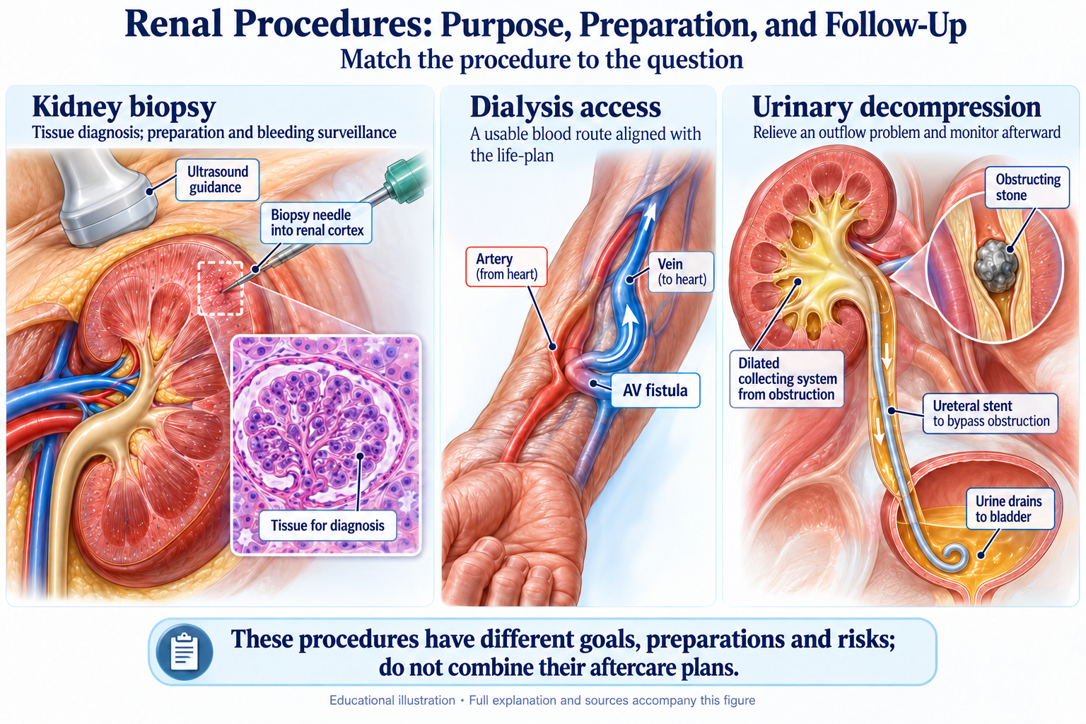
Kidney biopsy provides essential diagnostic and prognostic information for many glomerular diseases and unexplained kidney dysfunction. It is crucial for guiding specific immunosuppressive therapy.
Common Indications for Kidney Biopsy
- Nephrotic syndrome in adults
- Nephritic syndrome with reduced GFR
- Unexplained AKI or rapidly progressive glomerulonephritis (RPGN)
- Systemic disease with evidence of kidney involvement (e.g., lupus)
- Kidney transplant dysfunction or surveillance
Contraindications
Key Contraindications
- Uncorrectable bleeding disorder or coagulopathy
- Uncontrolled severe hypertension
- Single functioning native kidney (relative contraindication)
- Multiple bilateral cysts or small, end-stage kidneys
- Active urinary tract or perinephric infection
Procedure and Complications
Percutaneous kidney biopsy is typically performed using real-time ultrasound guidance with the patient in the prone position. After local anesthesia, a spring-loaded biopsy gun is used to obtain core specimens from the lower pole of the kidney. Post-procedure care involves several hours of bed rest and monitoring for complications, the most common of which is bleeding (perirenal hematoma or gross hematuria).
Vascular Access for Hemodialysis
Timely planning for durable hemodialysis access is critical for patients progressing towards ESRD.
The "Fistula First" Initiative
The preferred order of vascular access is: Arteriovenous Fistula (AVF) > Arteriovenous Graft (AVG) > Central Venous Catheter (CVC). This hierarchy is based on longevity and complication rates (thrombosis and infection).
- Arteriovenous Fistula (AVF): A direct surgical connection between an artery and a vein. It is the gold standard, offering the best long-term patency and lowest infection rates, but requires 2-3 months to mature.
- Arteriovenous Graft (AVG): A synthetic tube connects an artery and vein. It can be used sooner (2-3 weeks) but has higher rates of thrombosis and infection.
- Central Venous Catheter (CVC): A catheter placed into a large central vein. It provides immediate access but has the highest risk of infection and central venous stenosis. It should be considered a temporary bridge to permanent access whenever possible.
Peritoneal Dialysis (PD) Catheter Placement
A soft, flexible catheter is surgically placed into the peritoneal cavity. Proper catheter tip positioning in the pelvis is essential for optimal drainage. After placement, a "break-in" period of at least 2 weeks is typically required to allow the exit site to heal before full-volume exchanges are started.