Visual summary
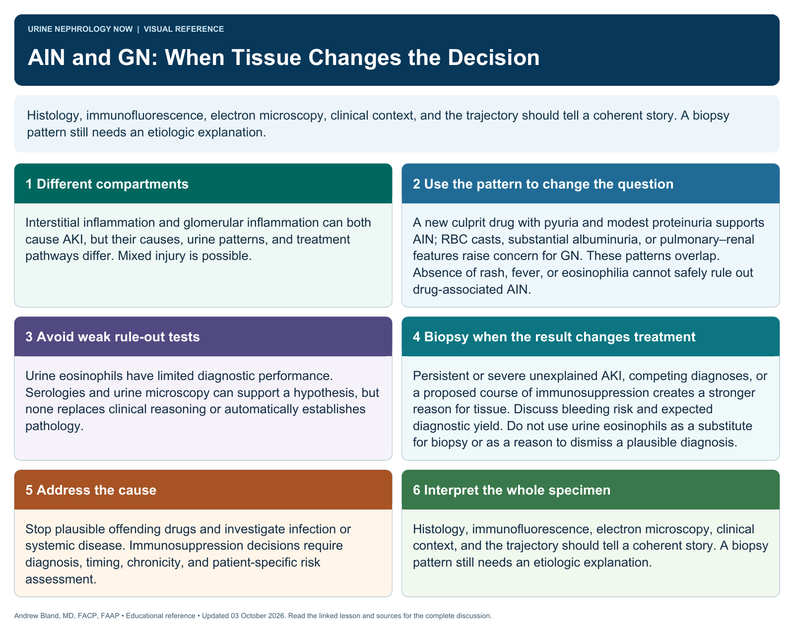
Histology, immunofluorescence, electron microscopy, clinical context, and the trajectory should tell a coherent story. A biopsy pattern still needs an etiologic explanation.
Text version
Different compartments
Interstitial inflammation and glomerular inflammation can both cause AKI, but their causes, urine patterns, and treatment pathways differ. Mixed injury is possible.
Use the pattern to change the question
A new culprit drug with pyuria and modest proteinuria supports AIN; RBC casts, substantial albuminuria, or pulmonary–renal features raise concern for GN. These patterns overlap. Absence of rash, fever, or eosinophilia cannot safely rule out drug-associated AIN.
Avoid weak rule-out tests
Urine eosinophils have limited diagnostic performance. Serologies and urine microscopy can support a hypothesis, but none replaces clinical reasoning or automatically establishes pathology.
Biopsy when the result changes treatment
Persistent or severe unexplained AKI, competing diagnoses, or a proposed course of immunosuppression creates a stronger reason for tissue. Discuss bleeding risk and expected diagnostic yield. Do not use urine eosinophils as a substitute for biopsy or as a reason to dismiss a plausible diagnosis.
Address the cause
Stop plausible offending drugs and investigate infection or systemic disease. Immunosuppression decisions require diagnosis, timing, chronicity, and patient-specific risk assessment.
Interpret the whole specimen
Histology, immunofluorescence, electron microscopy, clinical context, and the trajectory should tell a coherent story. A biopsy pattern still needs an etiologic explanation.