Visual summary
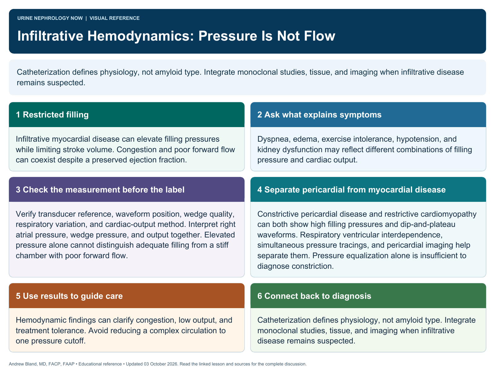
Catheterization defines physiology, not amyloid type. Integrate monoclonal studies, tissue, and imaging when infiltrative disease remains suspected.
Text version
Restricted filling
Infiltrative myocardial disease can elevate filling pressures while limiting stroke volume. Congestion and poor forward flow can coexist despite a preserved ejection fraction.
Ask what explains symptoms
Dyspnea, edema, exercise intolerance, hypotension, and kidney dysfunction may reflect different combinations of filling pressure and cardiac output.
Check the measurement before the label
Verify transducer reference, waveform position, wedge quality, respiratory variation, and cardiac-output method. Interpret right atrial pressure, wedge pressure, and output together. Elevated pressure alone cannot distinguish adequate filling from a stiff chamber with poor forward flow.
Separate pericardial from myocardial disease
Constrictive pericardial disease and restrictive cardiomyopathy can both show high filling pressures and dip-and-plateau waveforms. Respiratory ventricular interdependence, simultaneous pressure tracings, and pericardial imaging help separate them. Pressure equalization alone is insufficient to diagnose constriction.
Use results to guide care
Hemodynamic findings can clarify congestion, low output, and treatment tolerance. Avoid reducing a complex circulation to one pressure cutoff.
Connect back to diagnosis
Catheterization defines physiology, not amyloid type. Integrate monoclonal studies, tissue, and imaging when infiltrative disease remains suspected.