Visual summary
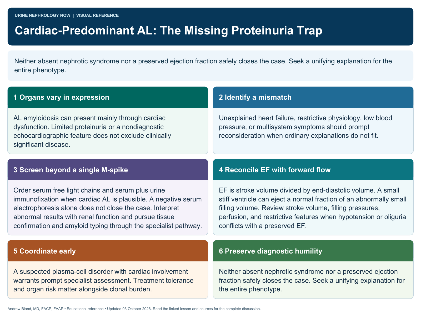
Neither absent nephrotic syndrome nor a preserved ejection fraction safely closes the case. Seek a unifying explanation for the entire phenotype.
Text version
Organs vary in expression
AL amyloidosis can present mainly through cardiac dysfunction. Limited proteinuria or a nondiagnostic echocardiographic feature does not exclude clinically significant disease.
Identify a mismatch
Unexplained heart failure, restrictive physiology, low blood pressure, or multisystem symptoms should prompt reconsideration when ordinary explanations do not fit.
Screen beyond a single M-spike
Order serum free light chains and serum plus urine immunofixation when cardiac AL is plausible. A negative serum electrophoresis alone does not close the case. Interpret abnormal results with renal function and pursue tissue confirmation and amyloid typing through the specialist pathway.
Reconcile EF with forward flow
EF is stroke volume divided by end-diastolic volume. A small stiff ventricle can eject a normal fraction of an abnormally small filling volume. Review stroke volume, filling pressures, perfusion, and restrictive features when hypotension or oliguria conflicts with a preserved EF.
Coordinate early
A suspected plasma-cell disorder with cardiac involvement warrants prompt specialist assessment. Treatment tolerance and organ risk matter alongside clonal burden.
Preserve diagnostic humility
Neither absent nephrotic syndrome nor a preserved ejection fraction safely closes the case. Seek a unifying explanation for the entire phenotype.