Visual summary
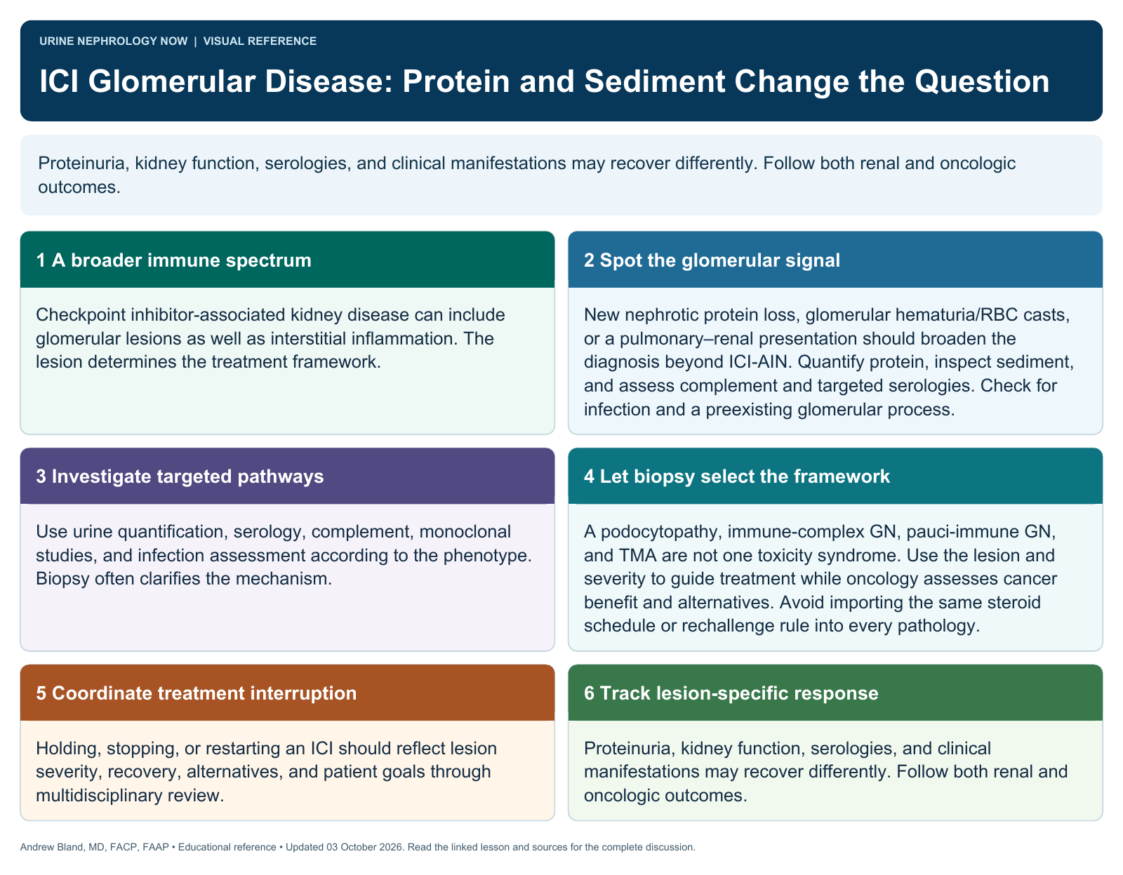
Proteinuria, kidney function, serologies, and clinical manifestations may recover differently. Follow both renal and oncologic outcomes.
Text version
A broader immune spectrum
Checkpoint inhibitor-associated kidney disease can include glomerular lesions as well as interstitial inflammation. The lesion determines the treatment framework.
Spot the glomerular signal
New nephrotic protein loss, glomerular hematuria/RBC casts, or a pulmonary–renal presentation should broaden the diagnosis beyond ICI-AIN. Quantify protein, inspect sediment, and assess complement and targeted serologies. Check for infection and a preexisting glomerular process.
Investigate targeted pathways
Use urine quantification, serology, complement, monoclonal studies, and infection assessment according to the phenotype. Biopsy often clarifies the mechanism.
Let biopsy select the framework
A podocytopathy, immune-complex GN, pauci-immune GN, and TMA are not one toxicity syndrome. Use the lesion and severity to guide treatment while oncology assesses cancer benefit and alternatives. Avoid importing the same steroid schedule or rechallenge rule into every pathology.
Coordinate treatment interruption
Holding, stopping, or restarting an ICI should reflect lesion severity, recovery, alternatives, and patient goals through multidisciplinary review.
Track lesion-specific response
Proteinuria, kidney function, serologies, and clinical manifestations may recover differently. Follow both renal and oncologic outcomes.