Visual summary
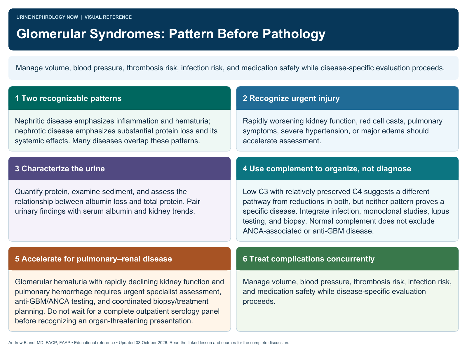
Manage volume, blood pressure, thrombosis risk, infection risk, and medication safety while disease-specific evaluation proceeds.
Text version
Two recognizable patterns
Nephritic disease emphasizes inflammation and hematuria; nephrotic disease emphasizes substantial protein loss and its systemic effects. Many diseases overlap these patterns.
Recognize urgent injury
Rapidly worsening kidney function, red cell casts, pulmonary symptoms, severe hypertension, or major edema should accelerate assessment.
Characterize the urine
Quantify protein, examine sediment, and assess the relationship between albumin loss and total protein. Pair urinary findings with serum albumin and kidney trends.
Use complement to organize, not diagnose
Low C3 with relatively preserved C4 suggests a different pathway from reductions in both, but neither pattern proves a specific disease. Integrate infection, monoclonal studies, lupus testing, and biopsy. Normal complement does not exclude ANCA-associated or anti-GBM disease.
Accelerate for pulmonary–renal disease
Glomerular hematuria with rapidly declining kidney function and pulmonary hemorrhage requires urgent specialist assessment, anti-GBM/ANCA testing, and coordinated biopsy/treatment planning. Do not wait for a complete outpatient serology panel before recognizing an organ-threatening presentation.
Treat complications concurrently
Manage volume, blood pressure, thrombosis risk, infection risk, and medication safety while disease-specific evaluation proceeds.