Visual summary
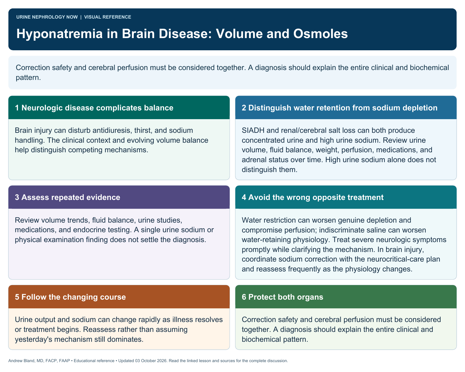
Correction safety and cerebral perfusion must be considered together. A diagnosis should explain the entire clinical and biochemical pattern.
Text version
Neurologic disease complicates balance
Brain injury can disturb antidiuresis, thirst, and sodium handling. The clinical context and evolving volume balance help distinguish competing mechanisms.
Distinguish water retention from sodium depletion
SIADH and renal/cerebral salt loss can both produce concentrated urine and high urine sodium. Review urine volume, fluid balance, weight, perfusion, medications, and adrenal status over time. High urine sodium alone does not distinguish them.
Assess repeated evidence
Review volume trends, fluid balance, urine studies, medications, and endocrine testing. A single urine sodium or physical examination finding does not settle the diagnosis.
Avoid the wrong opposite treatment
Water restriction can worsen genuine depletion and compromise perfusion; indiscriminate saline can worsen water-retaining physiology. Treat severe neurologic symptoms promptly while clarifying the mechanism. In brain injury, coordinate sodium correction with the neurocritical-care plan and reassess frequently as the physiology changes.
Follow the changing course
Urine output and sodium can change rapidly as illness resolves or treatment begins. Reassess rather than assuming yesterday's mechanism still dominates.
Protect both organs
Correction safety and cerebral perfusion must be considered together. A diagnosis should explain the entire clinical and biochemical pattern.
Supporting evidence
- Cerebral salt wasting: pathophysiology, diagnosis, and treatment.
- Differential diagnosis between syndrome of inappropriate antidiuretic hormone secretion and cerebral/renal salt wasting syndrome in children over 1 year: proposal for a simple algorithm.
- Risk of Overcorrection in Rapid Intermittent Bolus vs Slow Continuous Infusion Therapies of Hypertonic Saline for Patients With Symptomatic Hyponatremia: The SALSA Randomized Clinical Trial.