Visual summary
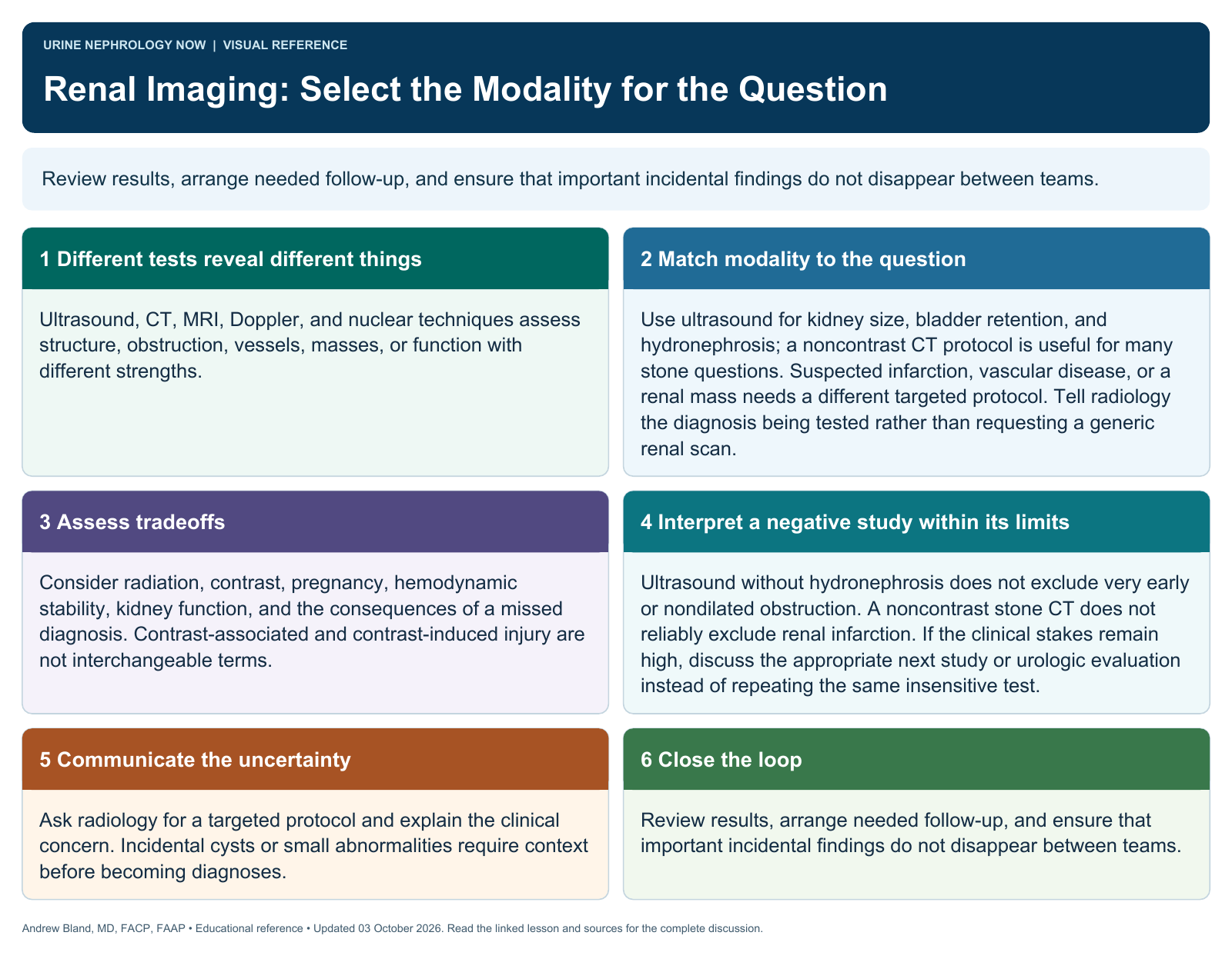
Review results, arrange needed follow-up, and ensure that important incidental findings do not disappear between teams.
Text version
Different tests reveal different things
Ultrasound, CT, MRI, Doppler, and nuclear techniques assess structure, obstruction, vessels, masses, or function with different strengths.
Start with a clinical question
Suspected obstruction, stones, renal mass, vascular disease, and unexplained AKI require different imaging strategies. Avoid ordering a modality without defining its purpose.
Assess tradeoffs
Consider radiation, contrast, pregnancy, hemodynamic stability, kidney function, and the consequences of a missed diagnosis. Contrast-associated and contrast-induced injury are not interchangeable terms.
Interpret with timing
Early obstruction and some nondilated processes may evade an initial scan. A negative result does not end investigation when clinical suspicion remains high.
Communicate the uncertainty
Ask radiology for a targeted protocol and explain the clinical concern. Incidental cysts or small abnormalities require context before becoming diagnoses.
Close the loop
Review results, arrange needed follow-up, and ensure that important incidental findings do not disappear between teams.
Continue learning
This graphic summarizes a topic. The full educational pages provide the broader discussion and references.