Visual summary
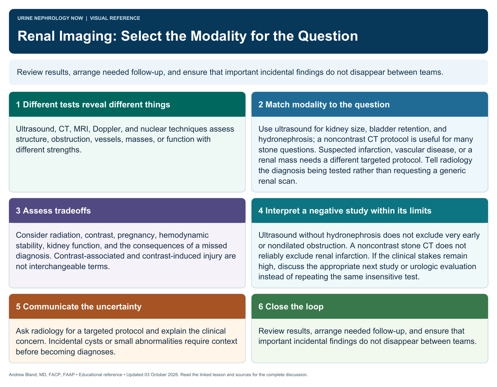
Review results, arrange needed follow-up, and ensure that important incidental findings do not disappear between teams.
Text version
Different tests reveal different things
Ultrasound, CT, MRI, Doppler, and nuclear techniques assess structure, obstruction, vessels, masses, or function with different strengths.
Match modality to the question
Use ultrasound for kidney size, bladder retention, and hydronephrosis; a noncontrast CT protocol is useful for many stone questions. Suspected infarction, vascular disease, or a renal mass needs a different targeted protocol. Tell radiology the diagnosis being tested rather than requesting a generic renal scan.
Assess tradeoffs
Consider radiation, contrast, pregnancy, hemodynamic stability, kidney function, and the consequences of a missed diagnosis. Contrast-associated and contrast-induced injury are not interchangeable terms.
Interpret a negative study within its limits
Ultrasound without hydronephrosis does not exclude very early or nondilated obstruction. A noncontrast stone CT does not reliably exclude renal infarction. If the clinical stakes remain high, discuss the appropriate next study or urologic evaluation instead of repeating the same insensitive test.
Communicate the uncertainty
Ask radiology for a targeted protocol and explain the clinical concern. Incidental cysts or small abnormalities require context before becoming diagnoses.
Close the loop
Review results, arrange needed follow-up, and ensure that important incidental findings do not disappear between teams.