Visual summary
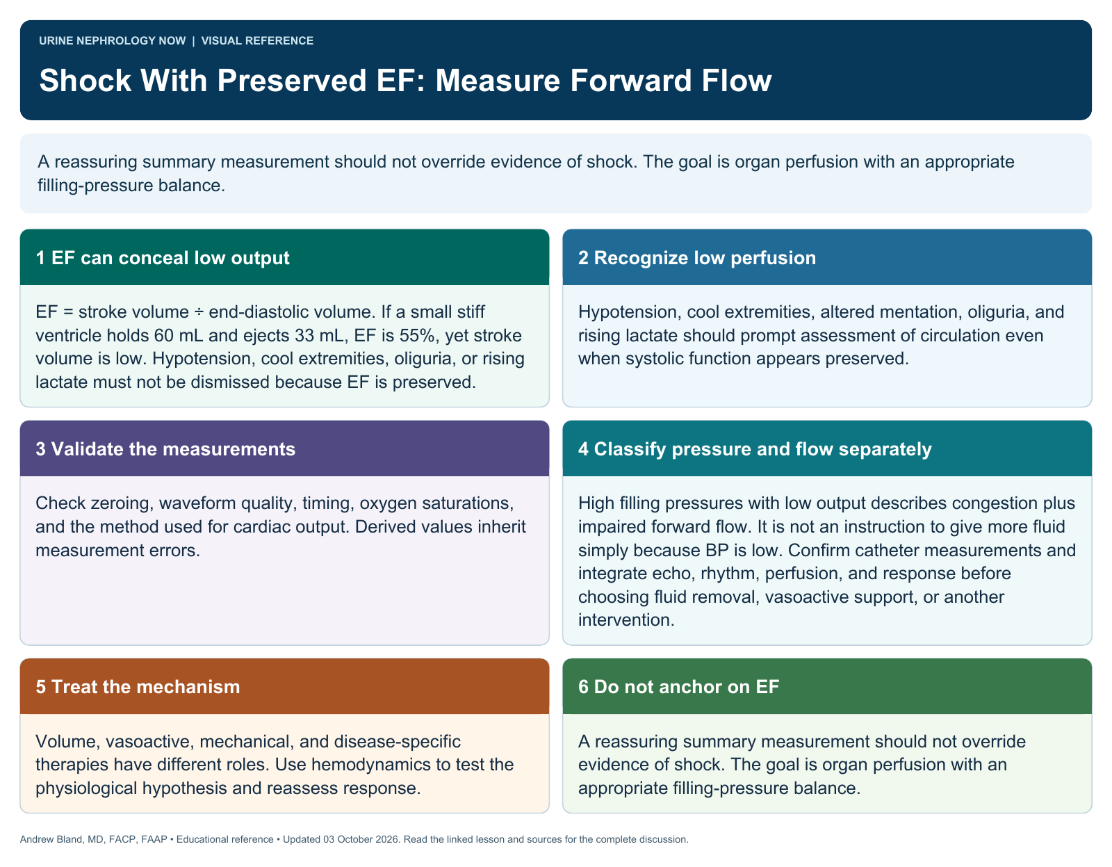
A reassuring summary measurement should not override evidence of shock. The goal is organ perfusion with an appropriate filling-pressure balance.
Text version
Ejection fraction is a fraction
A normal-looking ejection fraction can coexist with a small stroke volume and inadequate cardiac output. Shock is a perfusion syndrome, not an EF category.
Recognize low perfusion
Hypotension, cool extremities, altered mentation, oliguria, and rising lactate should prompt assessment of circulation even when systolic function appears preserved.
Validate the measurements
Check zeroing, waveform quality, timing, oxygen saturations, and the method used for cardiac output. Derived values inherit measurement errors.
Interpret the pattern
Combine right atrial pressure, pulmonary pressures, wedge pressure, output, and resistance with examination and imaging. Distinguish congestion from forward-flow limitation.
Treat the mechanism
Volume, vasoactive, mechanical, and disease-specific therapies have different roles. Use hemodynamics to test the physiological hypothesis and reassess response.
Do not anchor on EF
A reassuring summary measurement should not override evidence of shock. The goal is organ perfusion with an appropriate filling-pressure balance.
Continue learning
This graphic summarizes a topic. The full educational pages provide the broader discussion and references.