Visual summary
A reassuring summary measurement should not override evidence of shock. The goal is organ perfusion with an appropriate filling-pressure balance.
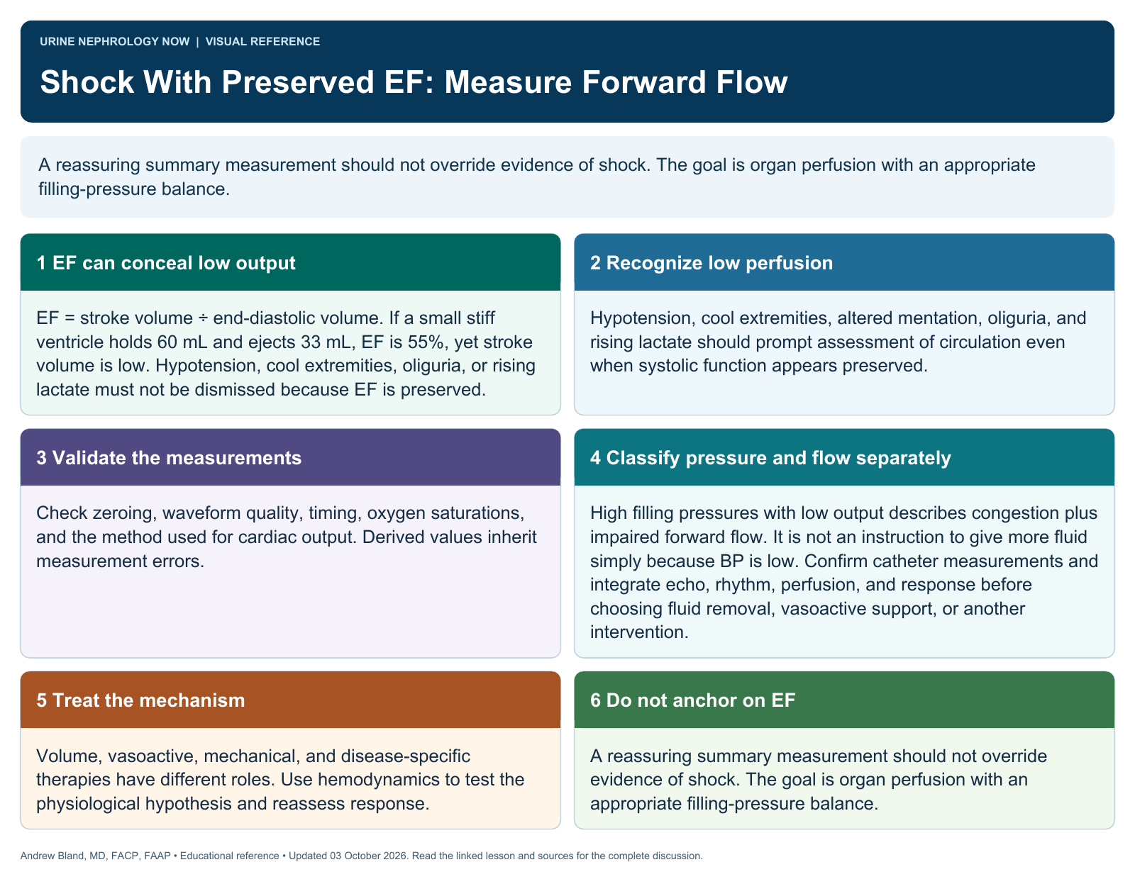
Text version
EF can conceal low output
EF = stroke volume ÷ end-diastolic volume. If a small stiff ventricle holds 60 mL and ejects 33 mL, EF is 55%, yet stroke volume is low. Hypotension, cool extremities, oliguria, or rising lactate must not be dismissed because EF is preserved.
Recognize low perfusion
Hypotension, cool extremities, altered mentation, oliguria, and rising lactate should prompt assessment of circulation even when systolic function appears preserved.
Validate the measurements
Check zeroing, waveform quality, timing, oxygen saturations, and the method used for cardiac output. Derived values inherit measurement errors.
Classify pressure and flow separately
High filling pressures with low output describes congestion plus impaired forward flow. It is not an instruction to give more fluid simply because BP is low. Confirm catheter measurements and integrate echo, rhythm, perfusion, and response before choosing fluid removal, vasoactive support, or another intervention.
Treat the mechanism
Volume, vasoactive, mechanical, and disease-specific therapies have different roles. Use hemodynamics to test the physiological hypothesis and reassess response.
Do not anchor on EF
A reassuring summary measurement should not override evidence of shock. The goal is organ perfusion with an appropriate filling-pressure balance.