Visual summary
Physiological plausibility and older trial data do not make every intervention equally proven. Reassess recurrence, tolerability, and the patient's ability to sustain the plan.
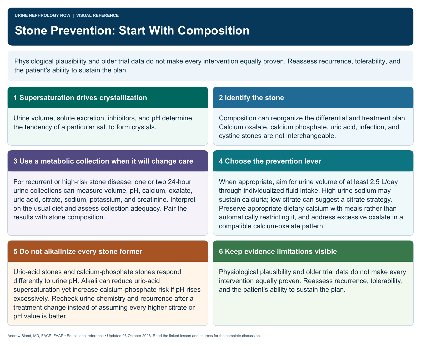
Text version
Supersaturation drives crystallization
Urine volume, solute excretion, inhibitors, and pH determine the tendency of a particular salt to form crystals.
Identify the stone
Composition can reorganize the differential and treatment plan. Calcium oxalate, calcium phosphate, uric acid, infection, and cystine stones are not interchangeable.
Use a metabolic collection when it will change care
For recurrent or high-risk stone disease, one or two 24-hour urine collections can measure volume, pH, calcium, oxalate, uric acid, citrate, sodium, potassium, and creatinine. Interpret on the usual diet and assess collection adequacy. Pair the results with stone composition.
Choose the prevention lever
When appropriate, aim for urine volume of at least 2.5 L/day through individualized fluid intake. High urine sodium may sustain calciuria; low citrate can suggest a citrate strategy. Preserve appropriate dietary calcium with meals rather than automatically restricting it, and address excessive oxalate in a compatible calcium-oxalate pattern.
Do not alkalinize every stone former
Uric-acid stones and calcium-phosphate stones respond differently to urine pH. Alkali can reduce uric-acid supersaturation yet increase calcium-phosphate risk if pH rises excessively. Recheck urine chemistry and recurrence after a treatment change instead of assuming every higher citrate or pH value is better.
Keep evidence limitations visible
Physiological plausibility and older trial data do not make every intervention equally proven. Reassess recurrence, tolerability, and the patient's ability to sustain the plan.