Visual summary
Identify loss versus shift, use urine and acid–base findings to localize the mechanism, and replace potassium with magnesium and kidney function in view.
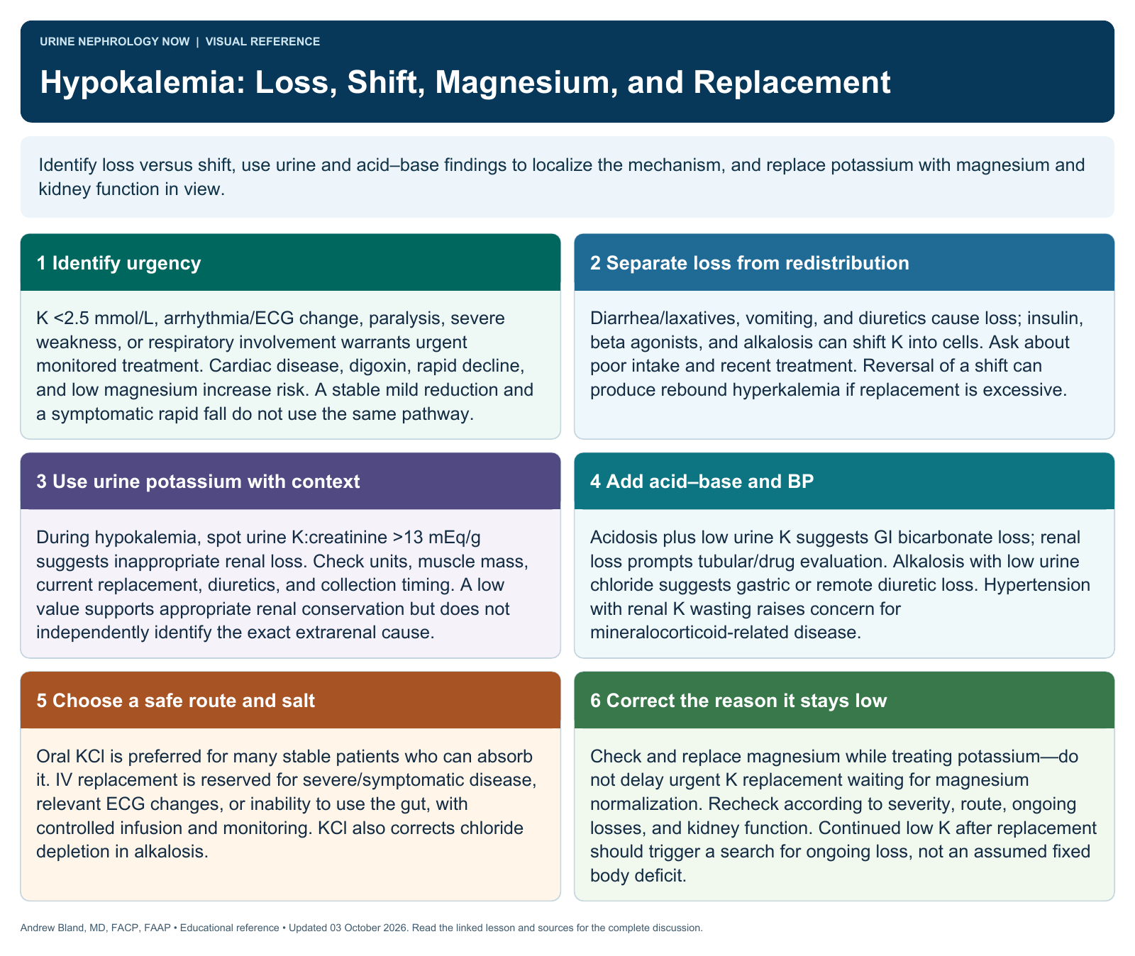
Text version
Identify urgency
K <2.5 mmol/L, arrhythmia/ECG change, paralysis, severe weakness, or respiratory involvement warrants urgent monitored treatment. Cardiac disease, digoxin, rapid decline, and low magnesium increase risk. A stable mild reduction and a symptomatic rapid fall do not use the same pathway.
Separate loss from redistribution
Diarrhea/laxatives, vomiting, and diuretics cause loss; insulin, beta agonists, and alkalosis can shift K into cells. Ask about poor intake and recent treatment. Reversal of a shift can produce rebound hyperkalemia if replacement is excessive.
Use urine potassium with context
During hypokalemia, spot urine K:creatinine >13 mEq/g suggests inappropriate renal loss. Check units, muscle mass, current replacement, diuretics, and collection timing. A low value supports appropriate renal conservation but does not independently identify the exact extrarenal cause.
Add acid–base and BP
Acidosis plus low urine K suggests GI bicarbonate loss; renal loss prompts tubular/drug evaluation. Alkalosis with low urine chloride suggests gastric or remote diuretic loss. Hypertension with renal K wasting raises concern for mineralocorticoid-related disease.
Choose a safe route and salt
Oral KCl is preferred for many stable patients who can absorb it. IV replacement is reserved for severe/symptomatic disease, relevant ECG changes, or inability to use the gut, with controlled infusion and monitoring. KCl also corrects chloride depletion in alkalosis.
Correct the reason it stays low
Check and replace magnesium while treating potassium—do not delay urgent K replacement waiting for magnesium normalization. Recheck according to severity, route, ongoing losses, and kidney function. Continued low K after replacement should trigger a search for ongoing loss, not an assumed fixed body deficit.