Visual summary
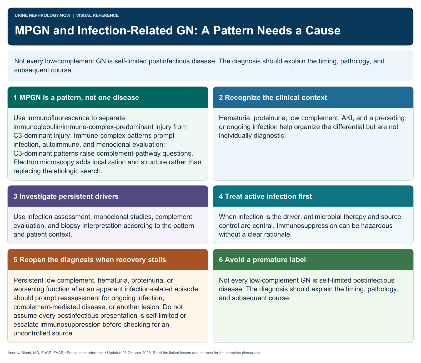
Not every low-complement GN is self-limited postinfectious disease. The diagnosis should explain the timing, pathology, and subsequent course.
Text version
MPGN is a pattern, not one disease
Use immunofluorescence to separate immunoglobulin/immune-complex-predominant injury from C3-dominant injury. Immune-complex patterns prompt infection, autoimmune, and monoclonal evaluation; C3-dominant patterns raise complement-pathway questions. Electron microscopy adds localization and structure rather than replacing the etiologic search.
Recognize the clinical context
Hematuria, proteinuria, low complement, AKI, and a preceding or ongoing infection help organize the differential but are not individually diagnostic.
Investigate persistent drivers
Use infection assessment, monoclonal studies, complement evaluation, and biopsy interpretation according to the pattern and patient context.
Treat active infection first
When infection is the driver, antimicrobial therapy and source control are central. Immunosuppression can be hazardous without a clear rationale.
Reopen the diagnosis when recovery stalls
Persistent low complement, hematuria, proteinuria, or worsening function after an apparent infection-related episode should prompt reassessment for ongoing infection, complement-mediated disease, or another lesion. Do not assume every postinfectious presentation is self-limited or escalate immunosuppression before checking for an uncontrolled source.
Avoid a premature label
Not every low-complement GN is self-limited postinfectious disease. The diagnosis should explain the timing, pathology, and subsequent course.