Visual summary
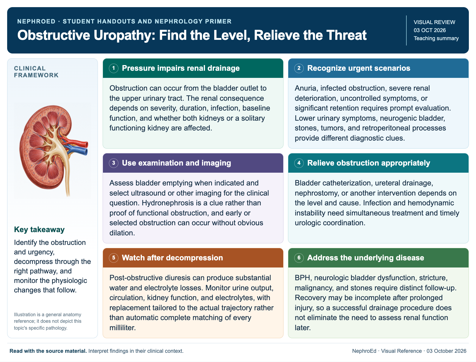
Identify the obstruction and urgency, decompress through the right pathway, and monitor the physiologic changes that follow.
Text version
Pressure impairs renal drainage
Obstruction can occur from the bladder outlet to the upper urinary tract. The renal consequence depends on severity, duration, infection, baseline function, and whether both kidneys or a solitary functioning kidney are affected.
Recognize urgent scenarios
Anuria, infected obstruction, severe renal deterioration, uncontrolled symptoms, or significant retention requires prompt evaluation. Lower urinary symptoms, neurogenic bladder, stones, tumors, and retroperitoneal processes provide different diagnostic clues.
Use examination and imaging
Assess bladder emptying when indicated and select ultrasound or other imaging for the clinical question. Hydronephrosis is a clue rather than proof of functional obstruction, and early or selected obstruction can occur without obvious dilation.
Relieve obstruction appropriately
Bladder catheterization, ureteral drainage, nephrostomy, or another intervention depends on the level and cause. Infection and hemodynamic instability need simultaneous treatment and timely urologic coordination.
Watch after decompression
Post-obstructive diuresis can produce substantial water and electrolyte losses. Monitor urine output, circulation, kidney function, and electrolytes, with replacement tailored to the actual trajectory rather than automatic complete matching of every milliliter.
Address the underlying disease
BPH, neurologic bladder dysfunction, stricture, malignancy, and stones require distinct follow-up. Recovery may be incomplete after prolonged injury, so a successful drainage procedure does not eliminate the need to assess renal function later.
Continue learning
This graphic summarizes a topic. The full educational pages provide the broader discussion and references.