Visual summary
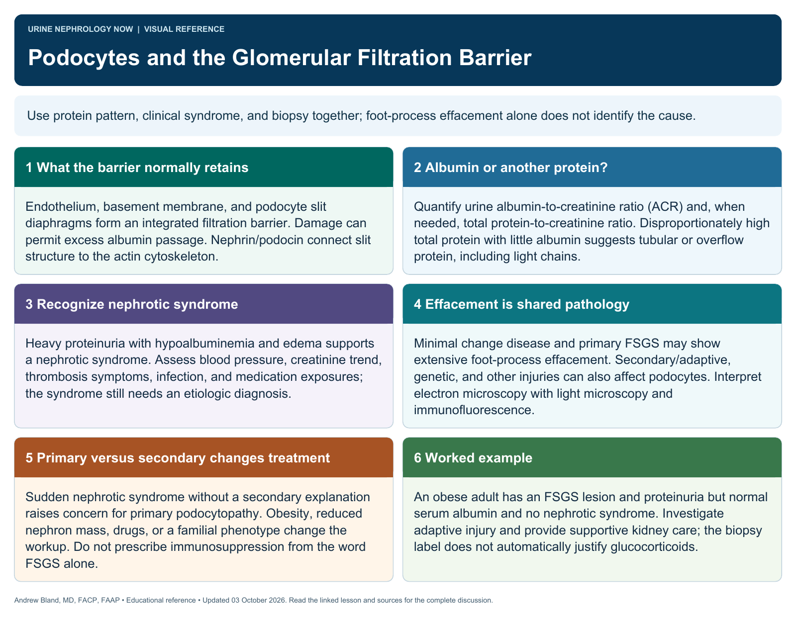
Podocyte biology explains important proteinuric patterns, but the disease diagnosis requires more than seeing foot-process effacement.
Text version
A coordinated filtration barrier
Fenestrated endothelium, the glomerular basement membrane, and podocyte architecture interact to restrict passage of blood cells and large proteins while allowing filtration. No single layer acts as an isolated sieve.
Foot processes and slit signaling
Podocyte foot processes and slit-diaphragm proteins connect structural organization to an actin cytoskeleton and signaling network. Genetic defects, immune injury, mechanical stress, or toxins can disturb that architecture.
Recognize the functional result
Proteinuria can indicate filtration-barrier dysfunction, but its mechanism may be glomerular, tubular, or overflow. Quantitative urine studies and clinical context help determine whether a podocyte-centered explanation is appropriate.
Interpret effacement carefully
Foot-process effacement on electron microscopy supports podocyte injury but is not unique to minimal change disease. The distribution, clinical syndrome, other biopsy findings, and potential secondary causes determine interpretation.
Link biology to treatment
Reducing glomerular stress and treating the identified disease can limit ongoing injury. Immune therapy, genetic evaluation, and supportive treatment serve different mechanisms; proteinuria alone does not justify a single universal regimen.
Check the model’s limits
Molecular pathways help organize learning, but a schematic does not prove which pathway drives an individual case. Avoid promising that every genetic association or experimental target already has a validated clinical treatment.
Continue learning
This graphic summarizes a topic. The full educational pages provide the broader discussion and references.