Visual summary
Use protein pattern, clinical syndrome, and biopsy together; foot-process effacement alone does not identify the cause.
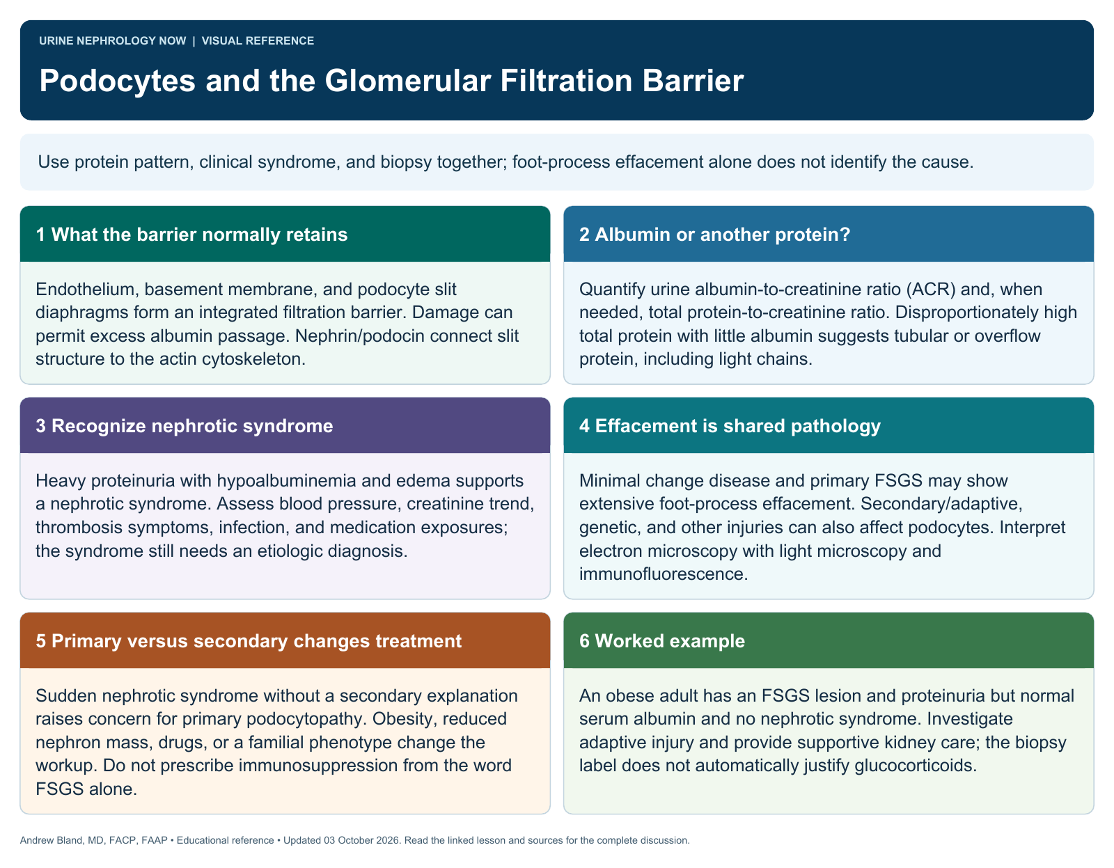
Text version
What the barrier normally retains
Endothelium, basement membrane, and podocyte slit diaphragms form an integrated filtration barrier. Damage can permit excess albumin passage. Nephrin/podocin connect slit structure to the actin cytoskeleton.
Albumin or another protein?
Quantify urine albumin-to-creatinine ratio (ACR) and, when needed, total protein-to-creatinine ratio. Disproportionately high total protein with little albumin suggests tubular or overflow protein, including light chains.
Recognize nephrotic syndrome
Heavy proteinuria with hypoalbuminemia and edema supports a nephrotic syndrome. Assess blood pressure, creatinine trend, thrombosis symptoms, infection, and medication exposures; the syndrome still needs an etiologic diagnosis.
Effacement is shared pathology
Minimal change disease and primary FSGS may show extensive foot-process effacement. Secondary/adaptive, genetic, and other injuries can also affect podocytes. Interpret electron microscopy with light microscopy and immunofluorescence.
Primary versus secondary changes treatment
Sudden nephrotic syndrome without a secondary explanation raises concern for primary podocytopathy. Obesity, reduced nephron mass, drugs, or a familial phenotype change the workup. Do not prescribe immunosuppression from the word FSGS alone.
Worked example
An obese adult has an FSGS lesion and proteinuria but normal serum albumin and no nephrotic syndrome. Investigate adaptive injury and provide supportive kidney care; the biopsy label does not automatically justify glucocorticoids.