Visual summary
Locate the structure, identify its transport function, and predict how injury or a medication changes the final urine.
Text version
Two linked pathways
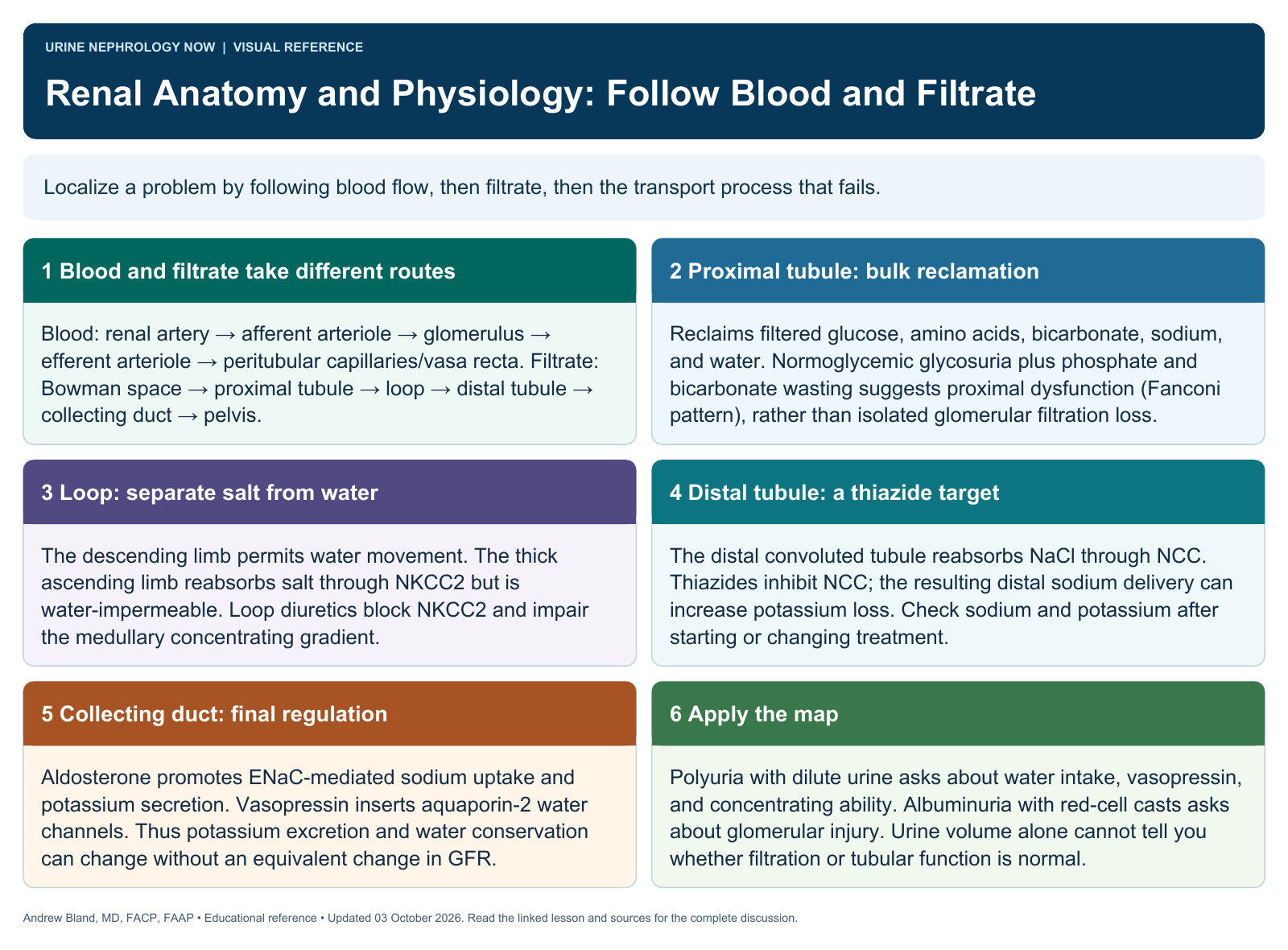
Blood enters the renal circulation and reaches glomerular capillaries, while filtered fluid enters Bowman space and moves through the nephron. Filtration and tubular processing together determine final urine composition.
Cortex and medulla
Glomeruli and much convoluted tubule occupy the cortex; loops and collecting structures extend into the medulla. Vascular arrangement helps preserve concentration gradients while delivering oxygen to metabolically active tissue.
Segment-specific transport
The proximal tubule reclaims large amounts of filtered solute and water. The loop builds concentration gradients, while distal segments and collecting ducts fine-tune electrolyte, water, and acid–base balance.
Control signals
Autoregulation, tubuloglomerular feedback, RAAS activity, vasopressin, and natriuretic signals coordinate filtration and excretion. These mechanisms respond to circulating volume, pressure, osmolality, and the body’s changing metabolic needs.
Connect structure to disease
Glomerular damage can cause proteinuria or hematuria; tubular injury can disturb electrolyte handling and concentrating ability. Vascular disease, interstitial inflammation, and obstruction affect different parts of the same integrated system.
Check your reasoning
Trace how a loop diuretic changes distal sodium delivery, or how vasopressin changes collecting-duct water permeability. Avoid treating filtration rate as equivalent to urine volume or overall tubular health.
Continue learning
This graphic summarizes a topic. The full educational pages provide the broader discussion and references.