Visual summary
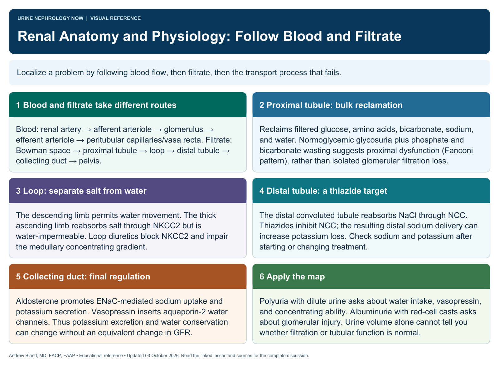
Localize a problem by following blood flow, then filtrate, then the transport process that fails.
Text version
Blood and filtrate take different routes
Blood: renal artery → afferent arteriole → glomerulus → efferent arteriole → peritubular capillaries/vasa recta. Filtrate: Bowman space → proximal tubule → loop → distal tubule → collecting duct → pelvis.
Proximal tubule: bulk reclamation
Reclaims filtered glucose, amino acids, bicarbonate, sodium, and water. Normoglycemic glycosuria plus phosphate and bicarbonate wasting suggests proximal dysfunction (Fanconi pattern), rather than isolated glomerular filtration loss.
Loop: separate salt from water
The descending limb permits water movement. The thick ascending limb reabsorbs salt through NKCC2 but is water-impermeable. Loop diuretics block NKCC2 and impair the medullary concentrating gradient.
Distal tubule: a thiazide target
The distal convoluted tubule reabsorbs NaCl through NCC. Thiazides inhibit NCC; the resulting distal sodium delivery can increase potassium loss. Check sodium and potassium after starting or changing treatment.
Collecting duct: final regulation
Aldosterone promotes ENaC-mediated sodium uptake and potassium secretion. Vasopressin inserts aquaporin-2 water channels. Thus potassium excretion and water conservation can change without an equivalent change in GFR.
Apply the map
Polyuria with dilute urine asks about water intake, vasopressin, and concentrating ability. Albuminuria with red-cell casts asks about glomerular injury. Urine volume alone cannot tell you whether filtration or tubular function is normal.