Visual summary
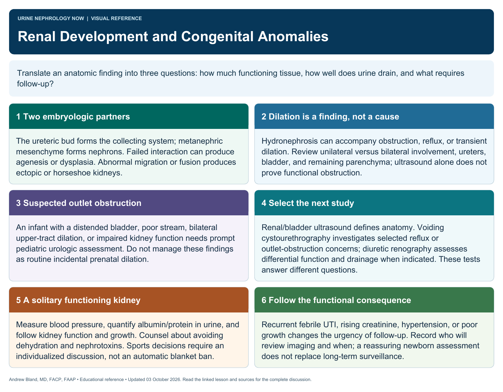
Translate an anatomic finding into three questions: how much functioning tissue, how well does urine drain, and what requires follow-up?
Text version
Two embryologic partners
The ureteric bud forms the collecting system; metanephric mesenchyme forms nephrons. Failed interaction can produce agenesis or dysplasia. Abnormal migration or fusion produces ectopic or horseshoe kidneys.
Dilation is a finding, not a cause
Hydronephrosis can accompany obstruction, reflux, or transient dilation. Review unilateral versus bilateral involvement, ureters, bladder, and remaining parenchyma; ultrasound alone does not prove functional obstruction.
Suspected outlet obstruction
An infant with a distended bladder, poor stream, bilateral upper-tract dilation, or impaired kidney function needs prompt pediatric urologic assessment. Do not manage these findings as routine incidental prenatal dilation.
Select the next study
Renal/bladder ultrasound defines anatomy. Voiding cystourethrography investigates selected reflux or outlet-obstruction concerns; diuretic renography assesses differential function and drainage when indicated. These tests answer different questions.
A solitary functioning kidney
Measure blood pressure, quantify albumin/protein in urine, and follow kidney function and growth. Counsel about avoiding dehydration and nephrotoxins. Sports decisions require an individualized discussion, not an automatic blanket ban.
Follow the functional consequence
Recurrent febrile UTI, rising creatinine, hypertension, or poor growth changes the urgency of follow-up. Record who will review imaging and when; a reassuring newborn assessment does not replace long-term surveillance.