Visual summary
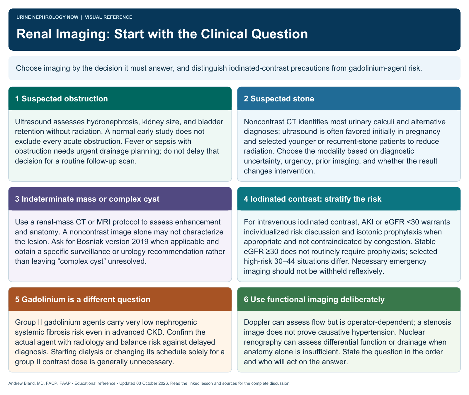
Choose the imaging study that best answers the question, with kidney precautions proportionate to the actual contrast and patient risk.
Text version
Different tests reveal different features
Ultrasound, CT, MRI, Doppler, and nuclear medicine studies offer complementary information about structure, obstruction, masses, blood flow, stones, and differential renal function. More detailed imaging is useful only when it answers a relevant question.
Recognize time-sensitive indications
Suspected infected obstruction, vascular catastrophe, serious trauma, or an important mass may require urgent imaging. Balance diagnostic urgency with the patient’s physiology instead of reflexively avoiding a test because kidney disease is present.
Select the initial modality
Ultrasound often helps evaluate hydronephrosis and structural disease; noncontrast CT is useful for many stone questions. Contrast-enhanced studies or MRI may be needed for characterization that an initial test cannot provide.
Plan contrast thoughtfully
Assess AKI, renal function, volume status, contrast type, route, and alternatives. Current risk assessment differs for iodinated contrast and gadolinium agents. Work with radiology to obtain an adequate study with appropriate precautions.
Use functional studies selectively
Duplex imaging and nuclear studies can answer focused vascular or functional questions, but test quality, operator dependence, and the clinical pretest probability influence interpretation. An anatomic abnormality does not automatically establish a treatable hemodynamic problem.
Avoid outdated blanket rules
Do not label all contrast agents equally nephrotoxic or all MRI contrast uniformly unsafe in CKD. Likewise, a low-risk designation is not a substitute for checking the specific agent, indication, and current guidance.
Self-check: State the clinical question before choosing ultrasound, CT, MRI, or a functional study, and explain whether contrast is needed to answer it.
Continue learning
This graphic summarizes a topic. The full educational pages provide the broader discussion and references.