Visual summary
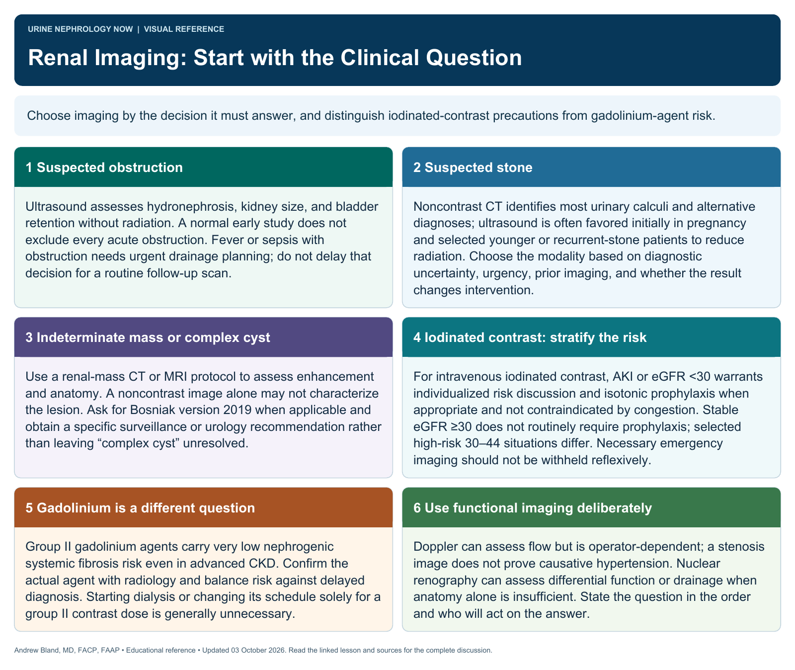
Choose imaging by the decision it must answer, and distinguish iodinated-contrast precautions from gadolinium-agent risk.
Text version
Suspected obstruction
Ultrasound assesses hydronephrosis, kidney size, and bladder retention without radiation. A normal early study does not exclude every acute obstruction. Fever or sepsis with obstruction needs urgent drainage planning; do not delay that decision for a routine follow-up scan.
Suspected stone
Noncontrast CT identifies most urinary calculi and alternative diagnoses; ultrasound is often favored initially in pregnancy and selected younger or recurrent-stone patients to reduce radiation. Choose the modality based on diagnostic uncertainty, urgency, prior imaging, and whether the result changes intervention.
Indeterminate mass or complex cyst
Use a renal-mass CT or MRI protocol to assess enhancement and anatomy. A noncontrast image alone may not characterize the lesion. Ask for Bosniak version 2019 when applicable and obtain a specific surveillance or urology recommendation rather than leaving “complex cyst” unresolved.
Iodinated contrast: stratify the risk
For intravenous iodinated contrast, AKI or eGFR <30 warrants individualized risk discussion and isotonic prophylaxis when appropriate and not contraindicated by congestion. Stable eGFR ≥30 does not routinely require prophylaxis; selected high-risk 30–44 situations differ. Necessary emergency imaging should not be withheld reflexively.
Gadolinium is a different question
Group II gadolinium agents carry very low nephrogenic systemic fibrosis risk even in advanced CKD. Confirm the actual agent with radiology and balance risk against delayed diagnosis. Starting dialysis or changing its schedule solely for a group II contrast dose is generally unnecessary.
Use functional imaging deliberately
Doppler can assess flow but is operator-dependent; a stenosis image does not prove causative hypertension. Nuclear renography can assess differential function or drainage when anatomy alone is insufficient. State the question in the order and who will act on the answer.