Visual summary
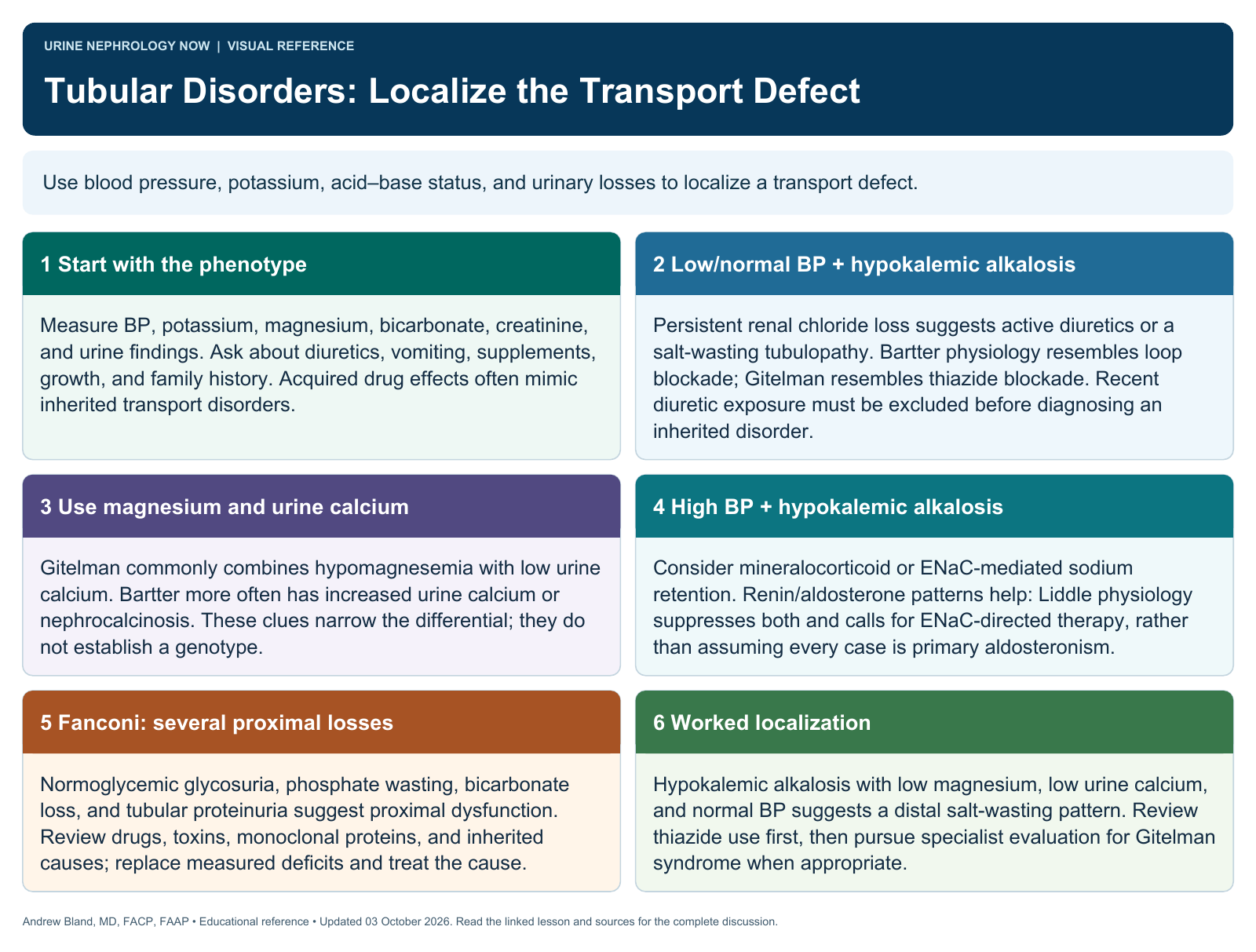
Use blood pressure, potassium, acid–base status, and urinary losses to localize a transport defect.
Text version
Start with the phenotype
Measure BP, potassium, magnesium, bicarbonate, creatinine, and urine findings. Ask about diuretics, vomiting, supplements, growth, and family history. Acquired drug effects often mimic inherited transport disorders.
Low/normal BP + hypokalemic alkalosis
Persistent renal chloride loss suggests active diuretics or a salt-wasting tubulopathy. Bartter physiology resembles loop blockade; Gitelman resembles thiazide blockade. Recent diuretic exposure must be excluded before diagnosing an inherited disorder.
Use magnesium and urine calcium
Gitelman commonly combines hypomagnesemia with low urine calcium. Bartter more often has increased urine calcium or nephrocalcinosis. These clues narrow the differential; they do not establish a genotype.
High BP + hypokalemic alkalosis
Consider mineralocorticoid or ENaC-mediated sodium retention. Renin/aldosterone patterns help: Liddle physiology suppresses both and calls for ENaC-directed therapy, rather than assuming every case is primary aldosteronism.
Fanconi: several proximal losses
Normoglycemic glycosuria, phosphate wasting, bicarbonate loss, and tubular proteinuria suggest proximal dysfunction. Review drugs, toxins, monoclonal proteins, and inherited causes; replace measured deficits and treat the cause.
Worked localization
Hypokalemic alkalosis with low magnesium, low urine calcium, and normal BP suggests a distal salt-wasting pattern. Review thiazide use first, then pursue specialist evaluation for Gitelman syndrome when appropriate.