Visual summary
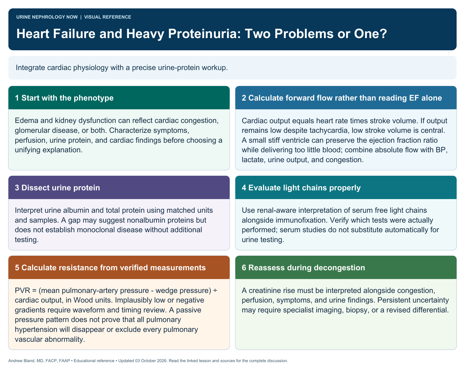
Integrate cardiac physiology with a precise urine-protein workup.
Text version
Start with the phenotype
Edema and kidney dysfunction can reflect cardiac congestion, glomerular disease, or both. Characterize symptoms, perfusion, urine protein, and cardiac findings before choosing a unifying explanation.
Read beyond ejection fraction
Assess ventricular geometry, diastolic function, right-heart findings, and forward output. Preserved or mildly reduced ejection fraction does not exclude severe hemodynamic disease.
Dissect urine protein
Interpret urine albumin and total protein using matched units and samples. A gap may suggest nonalbumin proteins but does not establish monoclonal disease without additional testing.
Evaluate light chains properly
Use renal-aware interpretation of serum free light chains alongside immunofixation. Verify which tests were actually performed; serum studies do not substitute automatically for urine testing.
Make hemodynamics coherent
When obtained, interpret filling pressures, cardiac output, and calculated resistance together. Check measurement quality before diagnosing restriction or assigning a pulmonary-hypertension subtype.
Reassess during decongestion
A creatinine rise must be interpreted alongside congestion, perfusion, symptoms, and urine findings. Persistent uncertainty may require specialist imaging, biopsy, or a revised differential.
Continue learning
This graphic summarizes a topic. The full educational pages provide the broader discussion and references.