Visual summary
Integrate cardiac physiology with a precise urine-protein workup.
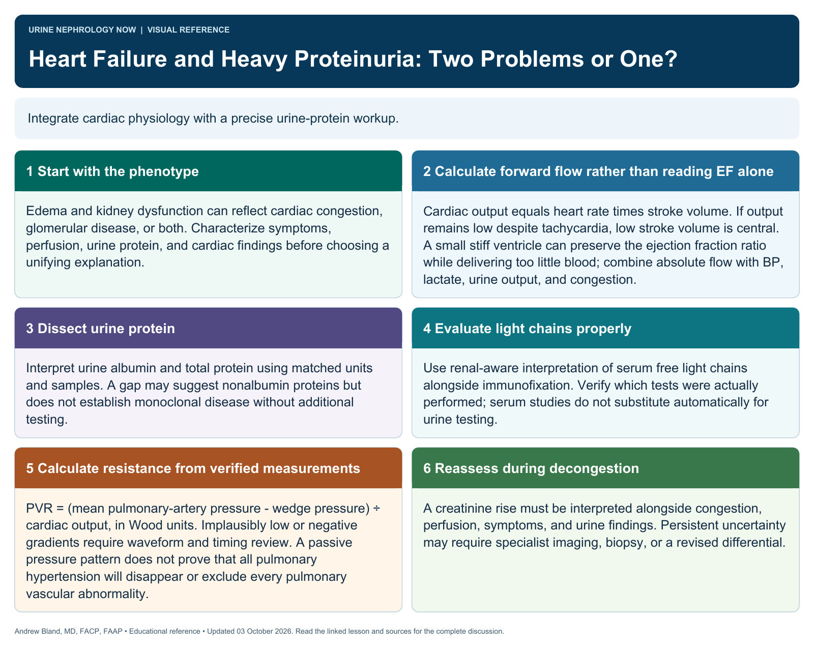
Text version
Start with the phenotype
Edema and kidney dysfunction can reflect cardiac congestion, glomerular disease, or both. Characterize symptoms, perfusion, urine protein, and cardiac findings before choosing a unifying explanation.
Calculate forward flow rather than reading EF alone
Cardiac output equals heart rate times stroke volume. If output remains low despite tachycardia, low stroke volume is central. A small stiff ventricle can preserve the ejection fraction ratio while delivering too little blood; combine absolute flow with BP, lactate, urine output, and congestion.
Dissect urine protein
Interpret urine albumin and total protein using matched units and samples. A gap may suggest nonalbumin proteins but does not establish monoclonal disease without additional testing.
Evaluate light chains properly
Use renal-aware interpretation of serum free light chains alongside immunofixation. Verify which tests were actually performed; serum studies do not substitute automatically for urine testing.
Calculate resistance from verified measurements
PVR = (mean pulmonary-artery pressure − wedge pressure) ÷ cardiac output, in Wood units. Implausibly low or negative gradients require waveform and timing review. A passive pressure pattern does not prove that all pulmonary hypertension will disappear or exclude every pulmonary vascular abnormality.
Reassess during decongestion
A creatinine rise must be interpreted alongside congestion, perfusion, symptoms, and urine findings. Persistent uncertainty may require specialist imaging, biopsy, or a revised differential.