Visual summary
Follow physiology across organ systems, then confirm the specific disease.
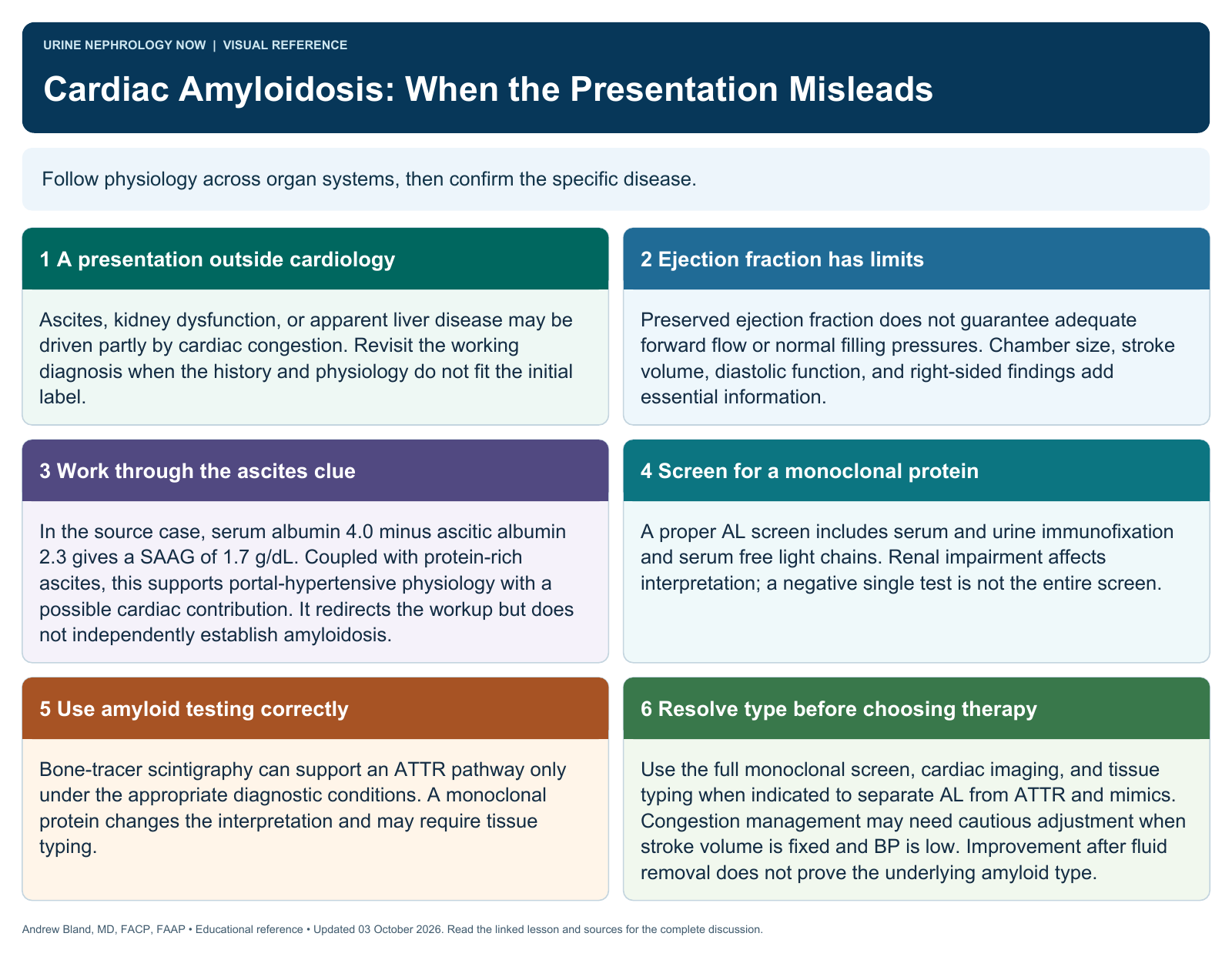
Text version
A presentation outside cardiology
Ascites, kidney dysfunction, or apparent liver disease may be driven partly by cardiac congestion. Revisit the working diagnosis when the history and physiology do not fit the initial label.
Ejection fraction has limits
Preserved ejection fraction does not guarantee adequate forward flow or normal filling pressures. Chamber size, stroke volume, diastolic function, and right-sided findings add essential information.
Use fluid and hemodynamic clues
Interpret ascitic fluid studies and, when indicated, right-heart catheterization in the full clinical context. No isolated pressure relationship or fluid-protein result proves infiltrative disease.
Screen for a monoclonal protein
A proper AL screen includes serum and urine immunofixation and serum free light chains. Renal impairment affects interpretation; a negative single test is not the entire screen.
Use amyloid testing correctly
Bone-tracer scintigraphy can support an ATTR pathway only under the appropriate diagnostic conditions. A monoclonal protein changes the interpretation and may require tissue typing.
Treat the established disease
AL and ATTR amyloidosis have different treatments. Coordinate typing, staging, congestion management, and shared goals of care. Avoid therapeutic decisions based only on a suggestive echocardiogram.
Continue learning
This graphic summarizes a topic. The full educational pages provide the broader discussion and references.