Visual summary
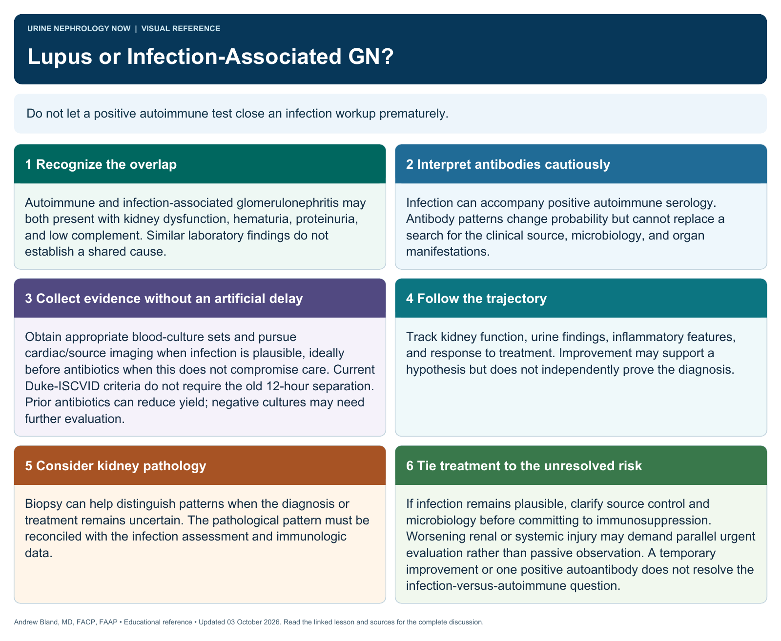
Do not let a positive autoimmune test close an infection workup prematurely.
Text version
Recognize the overlap
Autoimmune and infection-associated glomerulonephritis may both present with kidney dysfunction, hematuria, proteinuria, and low complement. Similar laboratory findings do not establish a shared cause.
Interpret antibodies cautiously
Infection can accompany positive autoimmune serology. Antibody patterns change probability but cannot replace a search for the clinical source, microbiology, and organ manifestations.
Collect evidence without an artificial delay
Obtain appropriate blood-culture sets and pursue cardiac/source imaging when infection is plausible, ideally before antibiotics when this does not compromise care. Current Duke-ISCVID criteria do not require the old 12-hour separation. Prior antibiotics can reduce yield; negative cultures may need further evaluation.
Follow the trajectory
Track kidney function, urine findings, inflammatory features, and response to treatment. Improvement may support a hypothesis but does not independently prove the diagnosis.
Consider kidney pathology
Biopsy can help distinguish patterns when the diagnosis or treatment remains uncertain. The pathological pattern must be reconciled with the infection assessment and immunologic data.
Tie treatment to the unresolved risk
If infection remains plausible, clarify source control and microbiology before committing to immunosuppression. Worsening renal or systemic injury may demand parallel urgent evaluation rather than passive observation. A temporary improvement or one positive autoantibody does not resolve the infection-versus-autoimmune question.