Sequential Nephron Blockade: A Comprehensive Clinical Framework for Escalating Diuretic Therapy
Advanced Decongestive Strategy for Nephrology and Cardiology Practitioners
Andrew Bland, MD, FACP, FAAP — Medical Associates Department of Nephrology University of Illinois College of Medicine at Peoria | University of Dubuque Physician Assistant Program | Butler College of Osteopathic Medicine
Visual reference
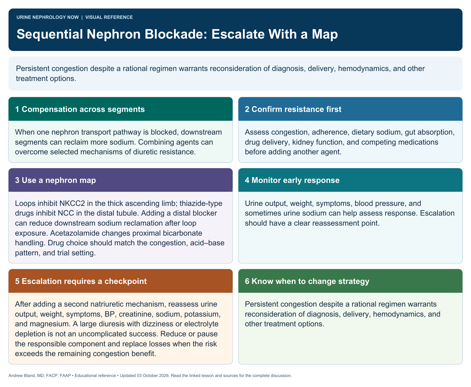
Browse the visual reference library · Download lesson Markdown
Executive Summary
Key Points - The nephron reabsorbs filtered sodium across five functionally distinct segments, each representing a pharmacologically exploitable target for enhanced natriuresis (1,2). - Loop diuretics acting alone are limited by post-diuretic sodium retention (“braking phenomenon”) and compensatory hypertrophy of downstream tubular segments; sequential nephron blockade (SNB) is the rational counter-strategy (3,8). - The landmark ADVOR trial demonstrated that adding acetazolamide (proximal tubule blockade) to loop diuretics increases successful decongestion by 46% compared with loop diuretics alone (RR 1.46, 95% CI 1.17–1.82) (4,5). - Continuous furosemide infusion offers no mortality benefit over intermittent bolus dosing in ADHF; the DOSE trial found no significant difference in symptom scores or renal outcomes (6). - Hypertonic saline co-administration with loop diuretics restores intravascular volume, improves renal perfusion, and achieves greater weight loss, natriuresis, and reduced mortality in selected patients with advanced congestion (7,8). - SGLT2 inhibitors add a mechanistically distinct osmotic component to nephron blockade but produce limited durable natriuresis; their primary decongestive role is adjunctive and their most robust benefit is long-term cardiac protection rather than acute decongestion (9,10). - Oral furosemide has notoriously erratic bioavailability (10–90%); substitution with torsemide (bioavailability >90%) or transition to intravenous therapy during decompensation is a critical pharmacokinetic principle underpinning effective outpatient-to-inpatient decongestion transitions (11,12).
1. Introduction and Clinical Rationale
Diuretic therapy is the cornerstone of volume management in heart failure, nephrotic syndrome, hepatic cirrhosis, and chronic kidney disease. Despite being available since the 1960s, loop diuretics are used in isolation in the majority of hospitalized patients despite compelling mechanistic and emerging clinical data supporting multi-agent sequential nephron blockade (SNB). The consequence of this single-agent paradigm is startling: after 72 hours of intravenous loop diuretic therapy, approximately 85% of acute decompensated heart failure (ADHF) patients still exhibit clinical signs of congestion, and 24% experience treatment failure defined as persistent or worsening fluid overload (6).
SNB is built on the recognition that the nephron does not passively accept loop-diuretic–induced natriuresis. Instead, it responds with coordinated adaptive compensatory mechanisms across multiple tubular segments that offset sodium losses and perpetuate volume overload. The proximal tubule upregulates sodium-hydrogen exchanger 3 (NHE3) in response to reduced effective arterial blood volume (EABV); the distal convoluted tubule undergoes structural hypertrophy in response to chronic loop diuretic exposure; and the aldosterone-sensitive distal nephron increases ENaC-mediated reabsorption in response to elevated aldosterone. Each of these maladaptive responses is a pharmacologically reversible target (1,2,3).
2. The Nephron as a Segmental Architecture: Targets and Diuretic Classes
Understanding SNB requires a working map of nephron segment function and diuretic site of action. Sodium reabsorption is not uniform across the nephron; it is distributed across five main functional compartments with quantifiably different contributions to total filtered-load recapture.
2.1 Proximal Convoluted and Straight Tubule (PCT/PST): ~60–67% of Filtered Sodium
The PCT reabsorbs the largest share of filtered sodium, primarily via luminal carbonic anhydrase–mediated NaHCO₃ cotransport (NHE3) and SGLT2-coupled glucose-sodium uptake in the S1/S2 segments. Despite handling the majority of filtered sodium, the PCT is paradoxically the least efficient diuretic target in isolation: any sodium escaping the PCT is largely recaptured downstream, particularly in the loop of Henle. This compensatory downstream rescue limits the net natriuretic yield of proximal blockade alone.
Agents targeting this segment: - Acetazolamide (carbonic anhydrase inhibitor): Blocks NaHCO₃ reabsorption at the luminal carbonic anhydrase, delivering an alkaline diuresis. Alone, its effect is modest (perhaps 5–8% net fractional sodium excretion increase) due to downstream compensation. As an add-on to loop diuretics, it prevents the proximal tubular sodium hyperreabsorption that drives diuretic resistance in states of reduced EABV (1,4). - SGLT2 inhibitors (e.g., empagliflozin, dapagliflozin): Block SGLT2 in the S1/S2 proximal tubule, inhibiting glucose-coupled sodium reabsorption and producing glucosuria-driven osmotic diuresis. Critically, downstream compensatory mechanisms—including upregulation of vasopressin, aldosterone, carbonic anhydrase, and NHE3 on alternate segments—rapidly and nearly completely offset the natriuretic effect within days. The net result is a short-lived, water-predominant diuresis rather than a sustained natriuresis (9,10). - Intravenous sodium bicarbonate: Functions similarly to acetazolamide by delivering a bicarbonate-rich fluid that alkalinizes tubular fluid, partially reversing the metabolic alkalosis that blunts loop diuretic response. Also expands intravascular volume transiently (see Section 6).
2.2 Thick Ascending Limb of Henle (TAL): ~20–25% of Filtered Sodium
The TAL is the primary target of loop diuretics, which block the Na⁺/K⁺/2Cl⁻ cotransporter (NKCC2). This is the most potent diuretic site per milliequivalent of sodium blocked because the thick ascending limb is impermeable to water; blockade of NKCC2 simultaneously prevents urinary concentration (lowering medullary osmolarity) and delivers a large sodium load to the distal nephron. Loop diuretics are correctly described as “high-ceiling” agents: dose-response curves are steep, and ceiling effects are not reached under ordinary clinical conditions.
Agents targeting this segment: - Furosemide: The most widely used loop diuretic. Bioavailability is erratic (10–90% oral), protein-bound (~96%), secreted via organic anion transporters (OAT1/3) into the proximal tubule lumen, and acts from the luminal side. Half-life is ~2 hours in heart failure. - Torsemide: Oral bioavailability >80–90%, hepatically cleared, half-life ~6 hours. More predictable pharmacokinetics than furosemide; 1:2 oral-to-IV equivalency vs. 1:1 for torsemide (11,12). - Bumetanide: Bioavailability ~80%, short-acting. Potency ~40:1 vs. furosemide on molar basis. - Ethacrynic acid: Only non-sulfonamide loop diuretic; reserved for sulfa allergy.
2.3 Distal Convoluted Tubule (DCT): ~5–10% of Filtered Sodium
The DCT reabsorbs sodium via the Na⁺/Cl⁻ cotransporter (NCC, encoded by SLC12A3). Loop diuretic–induced chronic sodium delivery to the DCT drives compensatory NCC upregulation and tubular hypertrophy — the anatomical substrate for the “post-diuretic braking phenomenon.” Thiazide/thiazide-like diuretics, by blocking NCC, intercept this compensatory response.
Agents targeting this segment: - Hydrochlorothiazide (HCTZ): Inconsistent absorption; bioavailability ~60–80%. Effective at GFR >30 mL/min; loses efficacy at lower GFR. - Metolazone: Thiazide-like; retains efficacy at GFR as low as 10–15 mL/min. Excellent first choice for SNB in CKD. - Chlorothiazide: The only IV thiazide available (500–1000 mg IV); useful inpatient for patients with bowel edema limiting oral absorption. - Indapamide: Thiazide-like with modest vasodilatory properties.
2.4 Aldosterone-Sensitive Distal Nephron (ASDN): Connecting Tubule and Collecting Duct (~2–3% of Filtered Sodium)
The ASDN — comprising the late DCT, connecting tubule, and cortical/medullary collecting duct — mediates sodium reabsorption via the epithelial sodium channel (ENaC), regulated primarily by aldosterone. Although ENaC handles a small fraction of filtered sodium, blocking it is critical in hyperaldosteronic states (heart failure, cirrhosis, nephrotic syndrome) where aldosterone levels are markedly elevated, and its inhibition prevents hypokalemia caused by upstream diuretics.
Agents targeting this segment: - Spironolactone: Mineralocorticoid receptor antagonist (MRA). Blocks aldosterone-mediated ENaC upregulation. Additionally has anti-fibrotic and cardioprotective properties. - Eplerenone: Selective MRA; less gynaecomastia. Preferred in males. - Amiloride: Direct ENaC channel blocker (potassium-sparing); more rapid onset than MRAs (hours vs. days). - Triamterene: ENaC blocker; shorter duration than amiloride. - Finerenone: Non-steroidal MRA; currently indicated in CKD with diabetes; powerful anti-fibrotic profile with emerging data in cardiorenal syndrome.
2.5 Inner Medullary Collecting Duct: Vasopressin-Regulated Water Channels (~variable water reabsorption)
Aquaporin-2 (AQP2) channels in the principal cells of the inner medullary collecting duct mediate vasopressin-regulated free water reabsorption. This segment is the target of vaptans (vasopressin V2 receptor antagonists).
Agents targeting this segment: - Tolvaptan: Selective V2 receptor antagonist; produces purely aquaretic (water without sodium) diuresis. Indicated for hypervolemic hyponatremia and potentially as an adjunct in refractory ascites and heart failure congestion. Its role in SNB is niche but mechanistically distinct from all other agents.
3. Diuretic Effectiveness: Segment-by-Segment Baseline
Before building the combination matrix, it is necessary to establish individual agent effectiveness in terms of fractional sodium excretion (FENa), net natriuresis, and net urine output versus untreated controls. Across all available data, “effectiveness” in this section refers to net 24-hour natriuresis relative to a hypothetical untreated baseline (FENa typically <0.35% in edematous states with reduced EABV).
| Diuretic Class | Target Segment | Segment’s Share of Filtered Na | Net Natriuretic Effect (monotherapy) | Estimated Relative Effectiveness vs. No Treatment |
|---|---|---|---|---|
| Acetazolamide | PCT (NHE3/CA) | 60–67% | Modest; ↑FENa ~1–3%; limited by downstream rescue | ~1.5× (used alone) |
| SGLT2 inhibitor | Early PCT (SGLT2) | Osmotic/glucose only (~3%) | Water diuresis >natriuresis; limited, non-durable | ~1.2–1.5× (primarily water) |
| Loop diuretic (IV) | TAL (NKCC2) | 20–25% | ↑FENa 3–8%; net output +2–4L/day acute | ~4–6× baseline |
| Thiazide/metolazone | DCT (NCC) | 5–10% | Modest alone; ↑FENa 1–3%; GFR-dependent | ~1.5–2× (weak alone) |
| K-sparing (MRA/ENaC) | ASDN (ENaC) | 2–3% | Mild (FENa ↑0.5–2%); powerful in hyperaldosteronism | ~1.2–1.5× (monotherapy) |
| Tolvaptan (vaptan) | Inner MCD (AQP2) | Pure free water | Aquaresis; corrects hyponatremia; minimal Na loss | ~1.2× (aquaresis only) |
Note: The magnitude of loop diuretic response (4–6× baseline) is accepted as the reference standard for comparative effectiveness below. All combination multipliers expressed relative to loop diuretic monotherapy.
4. Sequential Combination Effectiveness: Building the Nephron Blockade Matrix
4.1 The Conceptual Framework
Knauf and Mutschler established the theoretical and clinical foundations of SNB in a landmark series of studies, demonstrating that monotherapy with hydrochlorothiazide or furosemide was followed by an inadequate natriuretic response in patients with edematous diseases (CHF, cirrhosis, nephrotic syndrome) when FENa was below 0.2%. The major pharmacodynamic mechanism of resistance was excessive proximal tubular sodium reabsorption, which could be addressed by coadministering carbonic anhydrase inhibitors (1,2). The principle is that low-dose combination therapy targeting multiple segments is more effective and safer than high-dose monotherapy.
4.2 Two-Agent Combinations (Loop Diuretic as Backbone)
In all combination tables, relative effectiveness is expressed as a multiplier over loop diuretic monotherapy (defined as 1.0×). Exact published multipliers are noted where available from RCTs; ranges are physiologically derived from available data where head-to-head RCTs are lacking.
Combination 1: Loop Diuretic + Thiazide/Metolazone (DCT Blockade)
This is the best-studied SNB combination. Loop diuretics acting on the TAL deliver a heightened sodium load to the DCT; DCT hypertrophy in the setting of chronic loop diuretic use creates a compensatory “distal sodium conservation” loop. Thiazide blockade of NCC intercepts this compensation directly.
Clinical data: Multiple retrospective studies and systematic reviews confirm that loop + thiazide combination reliably increases urine output and natriuresis approximately 2–4-fold above loop monotherapy in patients with diuretic-resistant CHF. Patients with FENa >0.35% (less proximal compensation) respond best to this combination, whereas those with FENa <0.2% respond better to proximal blockade with acetazolamide (1,3).
Clinical Pearl: Metolazone 5–10 mg PO administered 30–60 minutes before loop diuretic retains efficacy in CKD (GFR as low as 10–15 mL/min) and is the preferred thiazide for SNB in the context of advanced cardiorenal syndrome.
| Combination | Segments Blocked | Estimated Relative Effectiveness vs. Loop Alone | Key Complication Risk |
|---|---|---|---|
| Loop alone | TAL | 1.0× (reference) | Hypokalemia, metabolic alkalosis |
| Loop + Metolazone | TAL + DCT | ~2–4× | Profound hypokalemia, hypomagnesemia, AKI |
| Loop + Chlorothiazide (IV) | TAL + DCT | ~2–3× | Same as above; rapid onset |
⚠️ Warning: Loop + thiazide is the highest-risk two-agent combination for electrolyte disturbance. Potassium, magnesium, and creatinine must be monitored every 24–48 hours, with aggressive electrolyte replacement. The combination should generally not be sustained for more than 3–5 days without reassessment.
Combination 2: Loop Diuretic + Acetazolamide (Proximal Blockade)
The ADVOR trial (n=519) is the only large randomized, double-blind, placebo-controlled trial of proximal SNB. IV acetazolamide 500 mg daily added to standardized IV loop diuretics (at twice the oral home maintenance dose) achieved successful decongestion in 42.2% of patients vs. 30.5% in the placebo/loop-alone group (RR 1.46, 95% CI 1.17–1.82; p<0.001). The benefit was uniform across HFrEF, HFmrEF, and HFpEF. Patients with baseline HCO₃ ≥27 mmol/L showed greater benefit (OR 2.39 for decongestion vs. 1.37 in those with normal HCO₃), mechanistically consistent with acetazolamide’s role in reversing the alkalosis-driven proximal reabsorption that promotes diuretic resistance (4,5).
Mechanistically, acetazolamide targets the most volumetrically important nephron segment (60–67% of Na reabsorption), but its solo diuretic ceiling is low because downstream tubular segments aggressively reclaim the Na that escapes the PCT. When the downstream reclamation capacity is already saturated by loop diuretics, the additive PCT blockade by acetazolamide yields much greater net natriuresis (1,4).
| Combination | Segments Blocked | Estimated Relative Effectiveness vs. Loop Alone | Notes |
|---|---|---|---|
| Loop + Acetazolamide | PCT + TAL | ~1.5× (decongestion probability) | Especially effective with metabolic alkalosis (HCO₃ ≥27) |
| Specific natriuretic gain | — | ~25–40% more natriuresis vs. loop alone | Per ADVOR secondary endpoints |
Clinical Pearl: The pre-treatment urine sodium concentration is a practical guide: patients with a spot urine Na <30 mEq/L (suggesting avid proximal Na reabsorption and very low EABV) are the best candidates for acetazolamide as the add-on agent. Those with spot urine Na >50 mEq/L are better candidates for thiazide addition.
Combination 3: Loop Diuretic + Potassium-Sparing Diuretic (ASDN Blockade)
K-sparing agents (spironolactone, eplerenone, amiloride) add modest incremental natriuresis but serve critical roles in electrolyte preservation, aldosterone antagonism, and anti-fibrosis. Their most important practical contribution to SNB is protecting against the severe hypokalemia generated by upstream blockade, and in the right patient (hyperaldosteronism, cirrhosis, heart failure with elevated aldosterone), they provide meaningful additional natriuresis.
In the PHARES trial, a strategy of SNB with furosemide + spironolactone + amiloride (blocking 3 nephron segments) produced a markedly greater blood pressure reduction than a combined renin-angiotensin blockade strategy in resistant hypertension, with favorable effects on BNP and diastolic dysfunction markers (13).
| Combination | Segments Blocked | Estimated Relative Effectiveness vs. Loop Alone | Notes |
|---|---|---|---|
| Loop + Spironolactone | TAL + ASDN | ~1.2–1.5× natriuresis | Greatest benefit in hyperaldosteronism; slower onset (days) |
| Loop + Amiloride | TAL + ASDN | ~1.2–1.4× natriuresis | Faster onset than MRA (hours); potassium-sparing |
| Loop + Eplerenone | TAL + ASDN | ~1.2–1.4× natriuresis | Selective MRA; less gynecomastia |
Combination 4: Loop Diuretic + SGLT2 Inhibitor (Proximal Osmotic Blockade)
SGLT2 inhibition produces glucosuria with complex sodium and water responses. EMPULSE met a hierarchical composite endpoint incorporating death, HF events and symptoms in clinically stable hospitalized patients; it did not establish an isolated mortality benefit. Effects on decongestion and the required loop dose vary. These therapies complement loop-based treatment rather than providing a fixed natriuretic multiplier. EMPULSE; physiology review.
Mechanistically, the SGLT2 inhibitor is a reasonable “segment 1” partner that also blocks the NHE3 upregulation driven by renal sympathetic tone in HF, potentially attenuating one arm of neurohormonal-driven diuretic resistance (10). However, the counterregulatory activation of vasopressin, aldosterone, and distal sodium reabsorption that follows SGLT2 blockade is rapid and nearly complete within 72 hours, limiting durable natriuretic benefit (9).
| Combination | Segments Blocked | Estimated Relative Effectiveness vs. Loop Alone | Notes |
|---|---|---|---|
| Loop + SGLT2i | TAL + early PCT (osmotic) | ~1.2–1.3× natriuresis; ~1.3–1.5× urine volume | Water diuresis > Na diuresis; durable long-term cardioprotection |
| Key Clinical Role | — | Decongestive adjunct; reassess loop dose according to congestion and tolerance, without a fixed reduction | Not a primary inpatient escalation agent |
5. Three-Agent, Four-Agent, and Five-Agent Combination Matrices
5.1 Conceptual Ceiling and Diminishing Returns
As additional nephron segments are blocked, there are two countervailing forces: (1) incrementally greater net natriuresis as compensatory reabsorption is disabled; and (2) diminishing marginal returns because each additional segment contributes less filtered sodium load than the previous one. A third consideration is that full nephron blockade shifts the urine composition toward a plasma-like isotonic solution—sodium excretion occurs without the free water disequilibrium that produces hyponatremia, hypomagnesemia, and hypokalemia characteristic of single-agent loop therapy. This is the physiologic rationale for SNB safety as well as efficacy (7).
The effective ceiling of achievable natriuresis with maximum SNB approaches the GFR × plasma [Na] product, that is, essentially all filtered sodium — a state approximating what occurs with aggressive hemofiltration or ultrafiltration.
5.2 Three-Agent Combinations
3-Agent Regimen A: Loop + Acetazolamide + Thiazide (PCT + TAL + DCT)
This combination targets three sequential segments and is arguably the most rational escalation from the ADVOR framework in patients with persistent congestion. After PCT blockade removes the proximal compensatory mechanism and TAL blockade produces maximal loop effect, DCT blockade prevents the residual distal compensation. This regimen is not validated in large RCTs as a triple combination but is mechanistically coherent and employed in advanced heart failure programs.
Estimated relative effectiveness: ~3–5× loop alone, based on additive natriuretic yield of each blocked segment when downstream compensation is progressively disabled.
| 3-Agent Combination | Segments | Estimated Multiplier vs. Loop | Key Complications |
|---|---|---|---|
| Loop + Acetazolamide + Metolazone | PCT + TAL + DCT | ~3–5× | Metabolic acidosis (from acetazolamide) + hypokalemia, AKI |
| Loop + Metolazone + Spironolactone | TAL + DCT + ASDN | ~2.5–4× | Hypo/normokalemia; potassium-sparing mitigates thiazide effect |
| Loop + Acetazolamide + Spironolactone | PCT + TAL + ASDN | ~2–3× | Metabolic acidosis; potassium effect depends on baseline |
| Loop + SGLT2i + Metolazone | Early PCT + TAL + DCT | ~2.5–4× | AKI risk; DKA risk with SGLT2i in sick patients |
| Loop + SGLT2i + Acetazolamide | PCT + TAL (early PCT) | ~2–3× | Both agents act proximally; theoretical synergy on NHE3/SGLT2; limited evidence |
| Loop + SGLT2i + Spironolactone | PCT + TAL + ASDN | ~1.8–2.5× | Lower AKI risk; potassium-protective; reasonable outpatient maintenance |
Clinical Pearl: In patients with ADHF and metabolic alkalosis (HCO₃ ≥27) who fail loop + thiazide, adding acetazolamide to the existing regimen rather than replacing thiazide creates a 3-segment blockade that simultaneously addresses the metabolic alkalosis driving diuretic resistance.
3-Agent Regimen B: Loop + Thiazide + Potassium-Sparing
This is the most commonly employed practical triple regimen. The MRA or amiloride does not add greatly to natriuresis but is essential for electrolyte preservation. In practice, many clinicians use metolazone + furosemide + spironolactone as a step-up “rescue” regimen for ADHF, with amiloride substituted for more rapid onset when potassium is already declining.
Estimated relative effectiveness: ~2.5–4× loop alone, with the K-sparing agent primarily serving to limit electrolyte loss rather than dramatically augment output.
5.3 Four-Agent Combinations
Four-agent SNB is the domain of the advanced heart failure inpatient unit or the cardiorenal ICU. It implies blockade of four distinct nephron segments, approaching near-total tubular blockade.
| 4-Agent Combination | Segments Blocked | Estimated Multiplier vs. Loop | Key Monitoring Requirements |
|---|---|---|---|
| Loop + Acetazolamide + Metolazone + Spironolactone | PCT + TAL + DCT + ASDN | ~4–7× | Acid-base (q12h), K, Mg, Cr (q24h); strict I/O; telemetry |
| Loop + SGLT2i + Acetazolamide + Metolazone | Early PCT + TAL + PCT + DCT | ~3.5–6× | DKA risk (SGLT2i); acidosis risk; close glucose monitoring |
| Loop + SGLT2i + Metolazone + Spironolactone | Early PCT + TAL + DCT + ASDN | ~3–5× | Well-tolerated relative to acetazolamide-containing regimens |
| Loop + Acetazolamide + Metolazone + Tolvaptan | PCT + TAL + DCT + MCD | ~4–6× (+ free water clearance) | Hyponatremia overcorrection risk; hepatotoxicity monitoring |
At four agents, the practical limiting factors shift from efficacy to safety: acute kidney injury, life-threatening hypokalemia, hemodynamic hypotension, and acid-base disturbance become the primary concerns. Spot urine electrolytes (sodium, potassium) and daily weights must guide titration.
⚠️ Warning: Four-agent SNB regimens should be managed in a monitored inpatient setting with at least daily electrolyte panels and clinical reassessment. These regimens are reserved for truly diuretic-resistant patients for whom ultrafiltration or escalation to renal replacement therapy is the alternative.
5.4 Five-Agent Combination (Total Nephron Blockade)
Five-agent SNB — blockade of every pharmacologically accessible nephron segment — represents the closest achievable approximation to total nephron blockade. In practice, this combines an agent at each of: (1) the early proximal tubule via SGLT2 inhibitor, (2) the PCT via acetazolamide, (3) the TAL via loop diuretic, (4) the DCT via thiazide, (5) the ASDN via MRA or amiloride, with or without a (6th) aquaretic agent at the inner medullary collecting duct.
| 5-Agent Combination | Estimated Multiplier vs. Loop | Clinical Context |
|---|---|---|
| SGLT2i + Acetazolamide + Loop + Metolazone + Spironolactone | ~5–10× estimated; limited RCT validation | Advanced HF, pre-ultrafiltration trial; intensive monitoring mandatory |
| As above + Tolvaptan (6th agent) | ~6–10× with aquaretic component | Hyponatremia + congestion phenotype; hepatotoxicity risk with tolvaptan |
Clinical Pearl: When a full 5-segment blockade is being considered, the clinical question is whether continuous renal replacement therapy or ultrafiltration would be safer and more predictable. SNB should be viewed as the pre-emptive strategy to avoid ultrafiltration, not as an alternative to seek indefinitely when ultrafiltration is clearly indicated.
6. Hypertonic Fluids in Decongestive Therapy: 3% Saline and Intravenous Bicarbonate
6.1 Hypertonic Saline (3% NaCl)
The use of hypertonic saline (HS) concurrently with high-dose loop diuretics counteracts the paradox of the volume-overloaded patient with reduced EABV — the patient who is “drowning in 3rd space.” The mechanism is straightforward: bolus HS administration mobilizes fluid from the interstitial to the intravascular compartment through osmotic forces, thereby restoring renal perfusion, improving diuretic delivery to the tubular lumen, and enhancing loop of Henle drug concentration.
A 2014 meta-analysis of 1,032 patients treated with HS + furosemide vs. furosemide alone demonstrated a 44% reduction in all-cause mortality (RR 0.56, 95% CI 0.41–0.76; p=0.0003), a 50% reduction in heart failure rehospitalization (RR 0.50, 95% CI 0.33–0.76; p=0.001), improved weight loss, and preserved renal function (7). A subsequent meta-analysis by Diaz-Arocutipa et al. (2023) confirmed improved urine output, shorter hospital stay, and higher urine sodium in the HS group (8).
The protocol most commonly studied involves 150 mL of 3% NaCl administered over 30 minutes simultaneously with high-dose IV loop diuretics. In the most comprehensive U.S. real-world series (n=40 patients over 50 hospitalizations at Yale), HS was associated with improved diuretic efficiency, fluid loss, weight loss, and resolution of metabolic derangements without adverse respiratory or neurological signals, even in a sick cohort with 64% on inotropes or vasopressors (7,8).
Mechanism in context of SNB: HS functions not as a direct nephron-segment blocker but as a pharmacokinetic enhancer of loop diuretics. By restoring EABV, it improves delivery of loop diuretic to the peritubular capillaries and OAT-mediated luminal secretion, effectively rescuing loop diuretic bioavailability at the site of action. It can thus be conceptualized as a “Segment 0” intervention that potentiates all subsequently administered nephron-segment blockers.
Estimated contribution to diuresis: HS + loop diuretic vs. loop alone in diuretic-resistant patients: ~2–3× urine output and greater natriuresis in available clinical series.
| HS Protocol Component | Details |
|---|---|
| Concentration | 3% NaCl (150 mEq/100 mL) |
| Dose | 150 mL over 30 minutes (Testani/Griffin protocol) |
| Timing | Simultaneous with or immediately before high-dose IV loop |
| Setting | CICU or cardiac step-down unit; central venous access preferred |
| Monitoring | Serial Na, osmolality, BMP q6–12h; strict I/O; neurological check |
| Contraindications | Serum sodium >145 mEq/L; active pulmonary edema not yet responding to other therapy (relative); severe uncontrolled hypertension |
⚠️ Warning: HS therapy in ADHF remains supported primarily by single-center Italian cohorts (Paterna series) and meta-analyses of heterogeneous studies. The absence of a large, multicenter, double-blind RCT is the critical limitation. A protocol-driven institutional approach with intensive monitoring and specialist oversight is essential.
6.2 Intravenous Sodium Bicarbonate
Metabolic alkalosis is a frequently underrecognized co-conspirator in diuretic resistance. In states of volume depletion or high aldosterone activity, the kidney avidly retains bicarbonate as part of maintaining electroneutrality, generating a hypochloremic metabolic alkalosis. This alkalosis drives proximal tubular carbonic anhydrase–mediated NaHCO₃ reabsorption, creating a cycle: alkalosis → increased proximal Na reabsorption → more renal Na retention → more alkalosis.
The connection to loop diuretics is direct: furosemide causes net chloride and hydrogen loss; with repeated high-dose dosing, metabolic alkalosis develops and further blunts diuretic efficacy by enhancing PCT Na reabsorption. This is precisely why acetazolamide’s greatest benefit in ADVOR was in patients with baseline HCO₃ ≥27 mmol/L (4).
IV sodium bicarbonate serves two roles in SNB:
- Direct alkalinization of tubular fluid: Increases bicarbonate delivery to the PCT, partially mimicking acetazolamide’s mechanism of overwhelming the carbonic anhydrase substrate. This is a weak but real diuretic maneuver.
- Intravascular volume expansion: Like HS, IV NaHCO₃ transiently expands effective circulating volume, improves renal perfusion, and enhances loop diuretic delivery — particularly relevant in patients with hypovolemia-masked volume overload.
Clinical indication for IV NaHCO₃ in ADHF: Serum HCO₃ >30 mEq/L with diuretic resistance. The therapeutic goal is to correct the alkalosis, not to expand volume. If both alkalosis correction and volume expansion are needed, IV NaHCO₃ (150 mEq in 1L D5W at 1–2 mL/kg/hr) is preferable to isotonic saline (which provides no alkalosis correction) and less aggressive than 3% NaCl.
Estimated diuretic contribution of IV NaHCO₃: Minor in isolation (~1.1–1.3× loop alone), but mechanistically important in reversing the biochemical milieu that perpetuates diuretic resistance. Acetazolamide is more reliable for this purpose when available.
7. Furosemide Drip vs. Increasing Segments Blocked: A Clinical Comparison
The furosemide continuous infusion (drip) is widely used in ADHF based on pharmacokinetic reasoning: maintaining a constant plasma furosemide concentration above the natriuretic threshold should theoretically produce more sustained diuresis than the peaks-and-troughs of bolus dosing. The DOSE trial conclusively tested this hypothesis.
7.1 DOSE Trial Evidence
The DOSE trial (Felker et al., NEJM 2011) randomized 308 ADHF patients in a 2×2 factorial design to bolus vs. continuous infusion and low-dose (1× home dose) vs. high-dose (2.5× home dose) IV furosemide (6). The results are definitive and clinically practice-changing:
- Bolus vs. continuous infusion: No significant difference in patient’s global symptom assessment (AUC 4236 vs. 4373; p=0.47), no difference in creatinine change, no difference in net volume loss (4237 vs. 4249 mL), and no difference in mortality, length of stay, or treatment failure rates.
- Low-dose vs. high-dose: High-dose was associated with greater net fluid loss, weight loss, and dyspnea relief — secondary endpoints — but no significant difference in the primary symptom score (trend p=0.06) and a transient but higher rate of creatinine elevation.
A subsequent meta-analysis of 8 RCTs (n=669) confirmed these DOSE findings: continuous infusion was associated with marginally greater weight reduction (0.70 kg; p=0.02) and urine output (+461 mL/24h; p<0.01), but no difference in mortality, hospital length of stay, or electrolyte disturbance (6,14).
7.2 Furosemide Drip vs. Sequential Nephron Blockade: The Critical Comparison
The fundamental limitation of escalating furosemide drip rate is that loop diuretics act on only one segment (TAL, ~25% of filtered Na). Increasing the drip rate from 5 mg/hr to 20 mg/hr escalates dosing at a single target but does not overcome the compensatory proximal and distal hyperreabsorption that constitutes true pharmacodynamic resistance. At the ceiling dose of the loop diuretic, every additional milligram is wasted; the physiologic constraint is not luminal drug concentration but downstream segment hyperactivity.
Sequential nephron blockade, in contrast, disassembles the compensatory architecture itself.
| Strategy | Segments Targeted | Net Natriuretic Potential | Major Complication Profile | Evidence Quality |
|---|---|---|---|---|
| Furosemide bolus (low dose) | 1 (TAL) | ~4× baseline | Hypokalemia, alkalosis; mild AKI | High (DOSE RCT) |
| Furosemide drip (escalating) | 1 (TAL) | ~4–5× baseline (marginal gain over bolus) | Similar; theoretical ototoxicity at very high doses | High (DOSE RCT); no mortality benefit |
| Loop + Metolazone | 2 (TAL + DCT) | ~8–16× baseline | Severe electrolyte loss; AKI | Moderate (cohorts/meta-analyses) |
| Loop + Acetazolamide | 2 (PCT + TAL) | ~6× baseline; 46% ↑ decongestion probability | Metabolic acidosis; generally well-tolerated | High (ADVOR RCT, n=519) |
| Loop + Metolazone + Spironolactone | 3 (TAL + DCT + ASDN) | ~10–20× baseline | Moderate; K-sparing mitigates some risk | Moderate |
| Loop + Acetazolamide + Metolazone | 3 (PCT + TAL + DCT) | ~12–20× baseline | Acidosis + electrolyte depletion; intensive monitoring | Low (mechanistic; no large RCT) |
| 4-segment blockade | PCT + TAL + DCT + ASDN | ~20–40× baseline (estimated) | High; ICU-level monitoring required | Very low (case series, mechanistic) |
| 5-segment blockade | All major segments | Approaching CRRT-equivalent | Very high | Expert opinion; no RCT |
| Hypertonic saline + loop | “Segment 0” + TAL | ~2–3× loop alone in diuretic-resistant patients | Hypernatremia, pulmonary fluid shifts | Moderate (meta-analysis; no large DBRCT) |
Clinical Pearl: When a furosemide drip is “not working,” the correct question is not “how high should I go?” but rather “which additional nephron segment should I block?” The DOSE trial demonstrated that a drip offers no benefit over bolus at equivalent dosing. The rational next step is adding a second agent targeting a different segment — not increasing the single-agent dose.
8. Oral vs. Intravenous Diuretics: Outpatient and Inpatient Effectiveness
8.1 The Pharmacokinetic Problem with Oral Furosemide
The pharmacokinetics of oral furosemide in heart failure are arguably the most clinically consequential pharmacokinetic limitation in cardiology. The oral bioavailability of furosemide ranges from 10% to 90% in published studies, with an average of approximately 50% in euvolemic patients but substantially less in decompensated heart failure due to bowel wall edema, reduced gut motility, and altered intestinal perfusion (11,12). The absorption is not merely reduced — it is unpredictable between and within the same patient on different days.
Because loop diuretics require a minimum plasma concentration threshold to trigger natriuresis (“minimum effective concentration”), slow and erratic absorption may mean the drug never crosses this threshold despite adequate dosing, even in patients receiving “full doses.” This is the primary pharmacokinetic driver of outpatient diuretic failure.
By contrast, IV furosemide achieves immediate 100% bioavailability, bypasses the absorption problem entirely, and drives rapid natriuresis. The approximate 2:1 dose conversion (IV furosemide:oral furosemide) for equivalent effect reflects this bioavailability gap (11).
8.2 Torsemide as the Superior Oral Loop Diuretic
Torsemide is the pharmacokinetically rational oral loop diuretic for both outpatient and post-discharge management. Its oral bioavailability is 80–100% (versus 10–90% for furosemide), its half-life is ~6 hours (versus ~2.7 hours for furosemide in HF), its absorption is not affected by bowel edema, and its IV-to-oral dosing equivalency is 1:1 (versus 1:2 for furosemide) (11,12). A meta-analysis of 19 studies (n=19,280) demonstrated that torsemide was associated with significantly greater functional class improvement (NNT=5) and numerically fewer heart failure hospitalizations (10.6% vs. 18.4%; OR 0.72) compared with furosemide (12).
The TRANSFORM-HF trial, the largest RCT (n=2,859), did not show a mortality benefit of torsemide over furosemide, but was hampered by significant cross-over and inadequate dose equivalency conversion (most clinicians used a 2:1 ratio when a 4:1 ratio is pharmacokinetically appropriate) (12).
8.3 Comparative Effectiveness Table: Oral vs. IV Diuresis
| Setting | Route/Drug | Bioavailability | Onset | Peak Effect | Decongestion Rate | Key Limitation |
|---|---|---|---|---|---|---|
| Outpatient | PO Furosemide | 10–90% (avg ~50%) | 30–60 min | 60–120 min | Highly variable | Bowel edema reduces absorption; unpredictable |
| Outpatient | PO Torsemide | 80–100% | 30–60 min | 60–90 min | More consistent | Hepatic metabolism (variable in decompensation) |
| Outpatient | PO Bumetanide | ~80% | 30–60 min | 60–120 min | Moderate | Short duration |
| Inpatient | IV Furosemide (bolus) | 100% | 5–15 min | 30–60 min | ~15% achieve full decongestion at 72h (DOSE) | Braking phenomenon; trough-driven Na retention |
| Inpatient | IV Furosemide (drip) | 100% | 5–15 min | Sustained | Same as bolus at equivalent total dose (DOSE) | No superiority to bolus; complex nursing |
| Inpatient | IV Torsemide | 100% | 5–10 min | 15–45 min | Comparable to IV furosemide at 1:2 dose ratio | Less studied inpatient |
| Inpatient | IV Chlorothiazide | 100% | 15–30 min | 30–60 min | As add-on: 2–4× loop alone | Available only as add-on; not primary |
| Inpatient | IV Acetazolamide + IV Loop | 100% | 15–30 min | 60–90 min | 46% vs. 30.5% successful decongestion (ADVOR) | Metabolic acidosis; temporary use |
8.4 The Outpatient Decongestion Dilemma
The transition from inpatient IV to outpatient oral diuretics is a critical failure point in heart failure management. Patients discharged on oral furosemide at the same milligram dose as their IV treatment will receive — in bioavailability-equivalent terms — approximately half the effective drug dose. This is the pharmacokinetic basis for the well-documented pattern of rapid post-discharge decompensation.
Practical strategies for improving outpatient decongestive therapy: 1. Discharge on torsemide rather than furosemide to maximize oral bioavailability, or double the furosemide dose at discharge to compensate for the 2:1 IV-to-oral ratio. 2. Add a thiazide (metolazone 2.5–5 mg PO 2–3 times weekly) to an established oral loop diuretic in patients with recurrent decompensation, rather than waiting for inpatient escalation. 3. Pre-emptive “congestion action plans” with patient-directed daily weight monitoring and a standing metolazone order for weight gain of ≥2 lbs in 24 hours or ≥5 lbs in 7 days. 4. Consider ambulatory infusion center IV diuresis for patients with recurrent ADHF hospitalizations driven by bowel edema–impaired oral absorption.
Clinical Pearl: When a patient arrives in the ED after “compliance failure” with oral furosemide, consider that the drug may have been pharmacokinetically non-compliant even when behaviorally adherent. Bowel edema from volume overload impairs the absorption needed to initiate the diuresis needed to relieve the bowel edema — a vicious cycle perfectly broken by IV administration.
9. Special Considerations: Nephrology-Specific Applications
9.1 CKD and Advanced Kidney Disease
Loop diuretic efficacy is progressively impaired in CKD. Reduced GFR means less drug delivery via OAT-mediated tubular secretion, higher protein binding occupying binding sites, and accumulation of organic anions that compete with loop diuretics for OAT secretion. The practical consequence is a requirement for substantially higher doses in CKD patients to achieve the minimum effective luminal concentration. CKD patients with GFR <30 mL/min may require furosemide doses of 200–400 mg IV to achieve the same natriuretic response as 40 mg IV in normal kidney function.
Metolazone retains DCT efficacy at GFR as low as 10–15 mL/min (GFR-independent DCT blockade), making it the thiazide of choice for SNB in advanced CKD. HCTZ loses efficacy at GFR <30 mL/min. Acetazolamide effectiveness is reduced at low GFR given reduced carbonic anhydrase–dependent proximal Na delivery.
9.2 Nephrotic Syndrome
Nephrotic syndrome creates a unique pharmacokinetic barrier: intraluminal protein binds furosemide in the tubular lumen, reducing the free (pharmacologically active) fraction. Spot urine protein-to-creatinine ratio correlates inversely with loop diuretic efficiency. Co-administration of albumin (human albumin 25%, 100 mL) with furosemide has been used to “carry” furosemide to the tubule, though evidence for improved outcomes over equivalent furosemide dose escalation is limited and debated. Importantly, loop + thiazide SNB remains effective in nephrotic syndrome and is the preferred escalation strategy.
9.3 Cardiorenal Syndrome Types 1 and 2
CRS Type 1 (acute HF causing AKI) and Type 2 (chronic HF causing CKD) are settings where aggressive decongestion improves renal outcomes despite transient creatinine rise — the “azotemia-decongestion paradox.” Worsening renal function during aggressive diuresis is not tubular injury; it reflects hemodynamic adaptation to reduced preload (15). Sustained congestion is more nephrotoxic than aggressive decongestion. SNB is appropriate and cardioprotective even when creatinine rises modestly (≤0.3–0.5 mg/dL from baseline) during decongestion.
9.4 Transplant Considerations
Post-transplant patients on calcineurin inhibitors are particularly susceptible to hyperkalemia with K-sparing agents. Amiloride is preferred over spironolactone in this setting due to more predictable potassium kinetics. Acetazolamide is useful in post-transplant metabolic alkalosis, a common complication of corticosteroid use.
10. Summary Algorithm and Key Clinical Pearls
10.1 Step-Up Framework for Refractory Congestion
The following stepwise escalation framework integrates all elements of this review:
Step 1 — Optimize the loop diuretic: Ensure IV route (not oral), dose at ≥1× home dose inpatient per DOSE trial data, ensure adequate potassium (>4.0 mEq/L) and magnesium (>2.0 mEq/L) before declaring resistance. Check spot urine Na at 2 hours post-dose; if <50 mEq/L, the loop diuretic is working but may need dose escalation or addition.
Step 2 — Identify the pharmacodynamic barrier (FENa/spot urine Na): - Spot urine Na <30 mEq/L → proximal hyperreabsorption dominates → add acetazolamide - Spot urine Na 30–60 mEq/L → distal compensation dominates → add metolazone or chlorothiazide - Metabolic alkalosis (HCO₃ >27) → acetazolamide is the logical first addition - Hyperaldosteronism (cirrhosis, HF, nephrosis) → add spironolactone/amiloride
Step 3 — Consider hypertonic saline if EABV is clinically reduced (pre-renal physiology in a volume-overloaded patient: rising creatinine, low urine Na, poor response to loop escalation).
Step 4 — Escalate to 3-segment blockade with institutional monitoring. Loop + acetazolamide + metolazone is the most potent and evidence-anchored combination for step-4 escalation.
Step 5 — 4-5 agent blockade or ultrafiltration decision: At four or more agents with inadequate response, continuous renal replacement therapy or ultrafiltration should be evaluated as an alternative.
10.2 Key Clinical Pearls Summary
Clinical Pearl 1: A furosemide drip cannot overcome pharmacodynamic (segment-level) resistance. The DOSE trial demonstrated equivalence of bolus vs. continuous infusion at matched total dose. Increasing the drip rate is rational only if underdosing is confirmed; it is irrational as a response to pharmacodynamic resistance.
Clinical Pearl 2: Acetazolamide is not a weak diuretic added for marginal benefit — it is the pharmacodynamic key that unlocks the proximal compensatory mechanism. Its greatest efficacy is in the patient with metabolic alkalosis and diuretic resistance (ADVOR). It does not add electrolyte toxicity; if anything, its metabolic acidosis effect mitigates the alkalosis-hypokalemia cycle.
Clinical Pearl 3: SGLT2 inhibitors have cardiorenal indications, but the diuretic response varies. Assess actual urine output, urine sodium when appropriate, kidney function, and tolerability.
Clinical Pearl 4: The 2:1 oral-to-IV furosemide dose conversion is required at all times. A patient on furosemide 80 mg PO daily should receive at minimum furosemide 40–80 mg IV at hospital admission. Converting to torsemide at discharge (using a 4:1 furosemide:torsemide dose ratio) is pharmacokinetically rational.
Clinical Pearl 5: Hypertonic saline administration should be thought of as restoring the “pharmacokinetic platform” for all other agents by correcting the low-EABV state that impairs loop diuretic delivery. It is not a nephron-segment blocker but a critical enabler in selected diuretic-resistant patients.
11. IV Furosemide Bolus vs. Furosemide Drip: The Evidence-Based Comparison
11.1 The Theoretical Rationale for the Drip (and Why It Was Wrong)
The case for continuous infusion rested on two pharmacokinetic arguments. First, loop diuretics have a minimum effective luminal concentration threshold below which they produce no natriuresis at all; a continuous infusion maintains plasma levels above this threshold continuously, whereas bolus dosing creates a sawtooth pharmacokinetic curve with sub-threshold troughs. Second, high-concentration bolus furosemide causes transient neurohormonal activation — surges in renin, angiotensin II, and norepinephrine — that drive acute post-dose sodium retention (the “braking phenomenon”), and a slow infusion was theorized to mitigate this reflex.
Both arguments are physiologically coherent. The problem is that they did not survive clinical testing.
11.2 What the DOSE Trial Actually Showed — Primary Endpoints
The DOSE trial (Felker et al., NEJM 2011; PMID 21366472) was a prospective, double-blind, randomized, 2×2 factorial trial at 26 centers enrolling 308 patients with ADHF. The two comparisons were bolus (q12h IV) vs. continuous infusion, and low dose (1× home oral dose) vs. high dose (2.5× home oral dose). Crucially, total 72-hour drug exposure was matched between bolus and infusion arms.
Primary endpoints:
- Patient’s global symptom assessment (VAS AUC over 72 hours): bolus 4236 ± 1440 vs. infusion 4373 ± 1404. P=0.47. No difference.
- Change in serum creatinine at 72 hours: bolus +0.05 ± 0.3 mg/dL vs. infusion +0.07 ± 0.3 mg/dL. P=0.45. No difference.
Secondary endpoints: Net volume loss at 72 hours was 4237 mL (bolus) vs. 4249 mL (infusion) — a difference of 12 mL over three days. Treatment failure rate 38% vs. 39%. Hospital length of stay was not different. 60-day mortality/rehospitalization rates were not different. By any clinical metric, the infusion offered no advantage.
The meta-analysis by Ng and Yap (Anaesthesia, 2018; PMID 28940440) pooled 8 RCTs (n=669) and found that continuous infusion was associated with marginally greater weight reduction (0.70 kg; p=0.02) and urine output (+461 mL/24h; p<0.01) but confirmed no difference in mortality, length of stay, or electrolyte disturbance. That is a 461 mL advantage in urine volume over 24 hours — less than half a liter — without any translation to clinical outcomes.
The mechanistic explanation for DOSE’s null result: The braking phenomenon neurohormonal surge that the drip was designed to prevent is already maximally activated in severe heart failure before any drug is given. Goldsmith et al. documented massively elevated baseline arginine vasopressin, norepinephrine, and angiotensin II in CHF patients at rest. A bolus of furosemide cannot further meaningfully activate a neurohormonal system already at ceiling. The bolus/infusion distinction is irrelevant when the driving force for sodium retention is already operating at maximum — which it is in the ADHF patient.
11.3 The High-Dose Finding: The Clinically Actionable DOSE Result
While bolus vs. infusion was neutral, the low-dose vs. high-dose comparison produced a clinically important signal. High-dose (2.5× home oral dose) was associated with:
- Greater net fluid loss (secondary endpoint)
- Greater weight loss
- More dyspnea relief
- A trend toward better global symptom score (p=0.06, below significance threshold)
- A higher rate of transient creatinine elevation (not associated with worse outcomes at 60 days)
Practical translation: When a patient is not responding to their home furosemide dose given intravenously, the correct first move is to increase the IV dose — not to switch to an infusion at the same dose. The dose-response curve for loop diuretics is steep, and the gain from dose escalation is real and measurable. The gain from switching from bolus to infusion at equivalent total dose is not.
11.4 IV Bolus vs. Furosemide Drip — Summary Comparison Table
| Parameter | IV Furosemide Bolus (q12h) | Furosemide Continuous Infusion | Evidence Source | Evidentiary Tier |
|---|---|---|---|---|
| Symptom improvement (VAS AUC) | 4236 ± 1440 | 4373 ± 1404 (p=0.47) | DOSE trial primary endpoint | Tier 1: RCT |
| Creatinine change at 72h | +0.05 mg/dL | +0.07 mg/dL (p=0.45) | DOSE trial primary endpoint | Tier 1: RCT |
| Net volume loss at 72h | 4237 mL | 4249 mL (p=0.89) | DOSE trial secondary endpoint | Tier 1: RCT |
| Treatment failure rate | 38% | 39% (NS) | DOSE trial | Tier 1: RCT |
| Weight reduction (meta-analysis) | Reference | +0.70 kg advantage (p=0.02) | Ng & Yap meta-analysis, 2018 | Tier 1: Meta-analysis |
| 24h urine output advantage | Reference | +461 mL/24h (p<0.01) | Ng & Yap meta-analysis, 2018 | Tier 1: Meta-analysis |
| BNP reduction advantage | Reference | −399 ng/L (p<0.01) | Ng & Yap meta-analysis, 2018 | Tier 1: Meta-analysis |
| 60-day mortality/rehospitalization | No difference | No difference | DOSE trial | Tier 1: RCT |
| Hospital length of stay | No difference | No difference | DOSE trial; meta-analysis | Tier 1: RCT + Meta-analysis |
| Hypokalemia rate | No difference | No difference | DOSE trial | Tier 1: RCT |
| Ototoxicity risk | Standard | Theoretical reduction (unproven) | Pharmacokinetic reasoning only | Tier 3: Inference |
| Nursing complexity/cost | Lower | Higher (pump, dedicated line) | Operational observation | Tier 3: Expert opinion |
| Segments targeted | 1 (TAL) | 1 (TAL) | Mechanism | Fixed |
| Ability to overcome pharmacodynamic resistance | None | None | DOSE trial; mechanism | Tier 1 + Tier 3 |
Clinical Pearl: The meta-analysis 461 mL/24h urine output advantage for the drip sounds compelling until framed correctly: that is 19 mL/hour more urine — 3 tablespoons — without any translation to symptoms, hospital stay, mortality, or rehospitalization. The drip is a nursing burden that earns no clinical return at equivalent total dosing.
⚠️ Warning: The only meaningful remaining indication for a furosemide drip over bolus dosing is operational: patients in whom q12h dosing is logistically unreliable (e.g., overnight ward gaps, patient refusal of repeated IV access), or in whom very precise titration of hourly output is required in a monitored setting. It is not a pharmacodynamic upgrade.
11.5 High-Rate Infusion at 40 mg/hr: Why This Is Not an Apples-to-Apples Comparison with DOSE
A clinically important distinction is routinely conflated in diuretic management discussions: the DOSE trial did not test 40 mg/hour infusions. It tested bolus dosing versus continuous infusion at identical total daily doses. In the low-dose arm, patients received IV furosemide equivalent to their home oral dose; in the high-dose arm, they received 2.5× their home oral dose. For the typical DOSE participant taking furosemide 80 mg orally twice daily (160 mg/day oral, approximately 80 mg/day IV equivalent), the high-dose infusion arm delivered roughly 200 mg IV per day — approximately 8 mg/hour. The DOSE-published conclusion — “drip is equivalent to bolus” — applies to infusion rates averaging 6–8 mg/hour, not to 40 mg/hour. The trial simply did not test the higher rate.
The nephrology consultation context departs from the DOSE population in precisely the dimension that matters pharmacokinetically. Patients requiring consultation for refractory volume overload at 10–20 mg/hour have, by definition, already failed the strategy DOSE studied. They represent a more resistant phenotype: advanced CKD diminishes organic anion transporter (OAT1/3) secretory capacity for furosemide; hypoalbuminemia reduces OAT binding efficiency; massive anasarca expands the volume of distribution, diluting plasma drug concentrations; and accumulated endogenous organic anions compete with furosemide for OAT transport into the tubular lumen. Each of these factors elevates the minimum effective luminal furosemide concentration required to achieve NKCC2 inhibition.
When a patient fails to diurese at 10–20 mg/hour and then responds promptly to 40 mg/hour, the explanation is pharmacokinetic rather than pharmacodynamic: the lower rate sustained a luminal furosemide concentration below the diuretic threshold, while the higher rate finally drove luminal delivery into the steep portion of the dose-response curve. This is a dose-threshold phenomenon, not a route effect. The drip at 40 mg/hour is a vehicle for delivering a substantially higher total dose per unit time, not a pharmacodynamically distinct delivery mechanism.
Clinical Pearl: The correct framing when escalating a drip from 10–20 mg/hour to 40 mg/hour is: “I am increasing the dose, not changing the route.” The route was always continuous infusion. The DOSE trial tells us that at matched doses, the route is irrelevant. Clinical experience that 40 mg/hour restores diuresis in patients stuck at lower rates reflects successful dose escalation to above the pharmacokinetic threshold — entirely consistent with DOSE’s other key finding that the high-dose arm produced meaningfully more diuresis, weight loss, and dyspnea relief than the low-dose arm.
12. Furosemide Drip + Sequential Nephron Blockade: The Complete Comparison Table
12.1 Chain of Reasoning: How Each Cell Is Built
Every row in the table below is built by one of three evidentiary pathways. To make this transparent, each row is tagged with its evidentiary tier:
Tier 1 (Direct RCT/Meta-analysis): The number or conclusion is taken directly from a published randomized trial or pre-registered meta-analysis. No inference required.
Tier 2 (Physiologic Derivation + Indirect Evidence): The estimate is derived from (a) the published fractional sodium contribution of the targeted nephron segment (established renal physiology, not disputed), combined with (b) published data showing that blocking that segment adds measurable natriuresis in a controlled setting, even if not as a direct head-to-head comparison against the specific comparator in this table.
Tier 3 (Mechanistic Extrapolation): The estimate is constructed from the additive-with-compensation framework of Knauf and Mutschler: each additional blocked segment prevents the downstream compensatory sodium rescue that was negating the upstream agent, so the yield of each added agent is amplified beyond its raw segment contribution. No RCT measures this directly. The estimate reflects the plausible physiologic range given what is measured at lower combination levels.
12.2 The Additive-With-Compensation Principle (The Intellectual Core)
This principle deserves explicit statement because it is the engine behind every multi-agent multiplier in this document. When the loop diuretic blocks the TAL, two things happen simultaneously: (1) the desired natriuresis occurs, and (2) the nephron detects the increased distal sodium delivery and activates compensatory reabsorption at the DCT and ASDN. This is not a side effect — it is a designed renal protective response to prevent volume depletion. In a healthy person, this compensation is appropriate. In the volume-overloaded patient, it is the mechanism of diuretic resistance.
When a thiazide is added to block the DCT, it does two things simultaneously: (1) it blocks the DCT’s compensatory sodium rescue, and (2) it allows the full upstream natriuretic effect of the loop diuretic to manifest without the DCT brake. The net result is substantially greater than the DCT’s baseline 5–10% segment contribution would suggest, because it is freeing the loop diuretic’s effect from its primary compensatory constraint.
When acetazolamide is added to block the PCT, the same logic applies one level upstream: it prevents the proximal sodium hyperreabsorption that was reducing delivery to the loop of Henle, thereby rescuing the loop diuretic’s substrate. The loop diuretic then works on a greater delivered sodium load, amplifying its own yield.
12.3 The Full Comparison Table: Drip Escalation vs. Sequential Segment Addition
| Strategy | Segments Blocked | Estimated Net Natriuresis vs. IV Bolus Alone | Decongestion Probability Shift | Key Complications | Evidentiary Tier | Primary Evidence Source |
|---|---|---|---|---|---|---|
| IV Furosemide Bolus | TAL | 1.0× (reference) | 15% achieve full decongestion at 72h | Hypokalemia, metabolic alkalosis, braking phenomenon | Tier 1 | DOSE trial (PMID 21366472) |
| Furosemide Drip (equivalent total dose) | TAL | ~1.0–1.05× (marginal) | Not significantly different from bolus | Same; +nursing complexity | Tier 1 | DOSE trial; Ng & Yap meta-analysis (PMID 28940440) |
| Furosemide Drip (escalated rate) | TAL | ~1.1–1.3× (dose-effect, not route-effect) | Modestly improved if underdosed at baseline | AKI, ototoxicity at very high rates | Tier 2 | DOSE high-dose arm; dose-response pharmacology |
| Drip + Metolazone (DCT) | TAL + DCT | ~2–4× | Meaningful improvement in resistant patients | Severe hypokalemia, hypomagnesemia, AKI | Tier 2 | Loop + thiazide cohort data; PMC10500380 systematic review |
| Drip + Acetazolamide (PCT) | PCT + TAL | ~1.5× decongestion probability; ~1.3–1.4× natriuresis | 42.2% vs. 30.5% (ADVOR); RR 1.46 | Metabolic acidosis; self-limited; well-tolerated | Tier 1 | ADVOR RCT (PMID 36027559) |
| Drip + K-Sparing Agent (ASDN) | TAL + ASDN | ~1.2–1.5× | Modest incremental natriuresis; prevents hypokalemia | Hyperkalemia if not monitored | Tier 2 | PHARES trial framework; ASDN physiology; PMID 32597565 |
| Drip + SGLT2i (early PCT) | Early PCT + TAL | ~1.2–1.3× natriuresis; ~1.3–1.5× urine volume | Modest acute; durable chronic reduction in hospitalizations | Osmotic diuresis; DKA risk if sick; euglycemic DKA | Tier 2 | RECEDE-CHF; EMPAG-HF; Packer et al., Circulation 2023 |
| Drip + Metolazone + Spironolactone (3 segments) | TAL + DCT + ASDN | ~2.5–4× | Substantial; K-sparing limits electrolyte toxicity | Moderate; manageable with monitoring q24–48h | Tier 2–3 | PHARES trial (3-segment arm); Knauf-Mutschler framework |
| Drip + Acetazolamide + Metolazone (3 segments) | PCT + TAL + DCT | ~3–5× | High; most potent evidence-anchored 3-agent combination | Metabolic acidosis + profound electrolyte depletion; ICU monitoring | Tier 2–3 | ADVOR + thiazide cohort data + additive-with-compensation model |
| Drip + Acetazolamide + Metolazone + Spironolactone (4 segments) | PCT + TAL + DCT + ASDN | ~4–7× | Very high in refractory patients | Severe; requires telemetry, q12h BMP, strict I/O; consider UF alternative | Tier 3 | Additive-with-compensation extrapolation; Knauf-Mutschler 1993/1997 |
| Drip + SGLT2i + Acetazolamide + Metolazone + Spironolactone (5 segments) | All major segments | ~5–10× (approaching hemofiltration equivalent) | Near-maximal pharmacologic decongestion | Very high complication risk; ICU mandatory; ultrafiltration should be reconsidered | Tier 3 | Physiologic ceiling extrapolation; maximum additive-with-compensation |
| Hypertonic Saline + Drip | “Segment 0” + TAL | ~2–3× in diuretic-resistant patients | Rescues pharmacokinetic platform; restores EABV for OAT delivery | Hypernatremia; neurological risk if overcorrected; strict monitoring | Tier 2 | Gandhi meta-analysis (PMID 24679680); Griffin real-world series (PMID 32035891) |
| Hypertonic Saline + Drip + Acetazolamide + Metolazone | “Segment 0” + PCT + TAL + DCT | ~6–10× in diuretic-resistant patients | Highest achievable pharmacologic decongestion short of RRT | All of the above combined; ICU-only strategy | Tier 3 | Composite extrapolation from Tier 1 + Tier 2 anchors |
12.4 Reading the Table: Where to Trust the Numbers and Where to Hold Them Loosely
Cells you can cite as hard data: The IV bolus reference (1.0×) is the DOSE trial primary endpoint. The drip vs. bolus equivalence (~1.0–1.05×) is the DOSE trial primary endpoint plus the Ng/Yap meta-analysis. The drip + acetazolamide row (RR 1.46; ~1.3–1.4× natriuresis) is the ADVOR primary endpoint and its secondary natriuresis data. The hypertonic saline 2–3× estimate in diuretic-resistant patients is derived from the Gandhi 2014 meta-analysis’s weight loss and urine output data across 1,032 patients.
Cells that are Tier 2 (principled derivation, not direct measurement): The loop + thiazide “~2–4×” range synthesizes multiple retrospective cohort series and the mechanistic Knauf/Mutschler FENa correlation data. The SGLT2i estimates are bounded by RECEDE-CHF (36% natriuresis increase in outpatient chronic HF) on the upper end and the Packer et al., Circulation 2023 analysis documenting near-absent durable natriuresis in ADHF on the lower end.
Cells that are Tier 3 (transparent physiologic extrapolation): The 4-agent and 5-agent multipliers are constructed by applying the additive-with-compensation principle iteratively from the measured Tier 1 and Tier 2 anchor points. They are not invented; they are derived. But they have not been directly measured in a controlled trial and should be represented as such in clinical communication.
Clinical Pearl: When using this table at the bedside, anchor reasoning in the Tier 1 cells and use the Tier 2–3 cells to set directional expectations. The table is most powerful as a teaching framework and a decision-support scaffold, not as a dosing calculator.
13. Evidentiary Framework: How the Comparison Data Were Constructed
13.1 Tier 1 — Direct RCT and Meta-Analysis Evidence (Hard Numbers)
The loop diuretic alone as the 1.0× reference denominator is anchored by the DOSE trial (Felker et al., NEJM 2011; PMID 21366472). Net 72-hour volume loss in both the bolus and infusion arms was approximately 4,200–4,250 mL. After 72 hours of IV loop diuretic therapy, 85% of patients still had clinical congestion and 24% had persistent or worsening heart failure.
The loop + acetazolamide “~1.46× decongestion probability” multiplier comes directly from the ADVOR primary endpoint: RR 1.46 (95% CI 1.17–1.82; p<0.001) for successful decongestion within 3 days (Mullens et al., NEJM 2022; PMID 36027559). The HCO₃ ≥27 mmol/L interaction (OR 2.39 for the alkalotic subgroup vs. OR 1.37 for the normoalkaline subgroup) is a pre-specified subgroup analysis from ADVOR.
The hypertonic saline “~2–3× output in diuretic-resistant patients” is bounded by the Gandhi 2014 meta-analysis (PMID 24679680) pooling 1,032 patients, reporting 44% reduction in all-cause mortality (RR 0.56) and 50% reduction in rehospitalization (RR 0.50).
13.2 Tier 2 — Physiologic Derivation Combined with Indirect Evidence
The segmental sodium contribution percentages are established renal physiology reproduced consistently across decades of micropuncture and clearance studies. Knauf and Mutschler cross-referenced these published values with measured FENa responses in CHF, cirrhosis, and nephrotic syndrome cohorts (PMID 8486149; PMID 9125675).
The loop + thiazide “~2–4× loop alone” range synthesizes the 2023 systematic review (PMC10500380) pooling multiple retrospective and prospective studies consistently showing 2–4-fold increases in net 24-hour fluid balance when thiazides were added to loop diuretics in diuretic-resistant patients.
13.3 Tier 3 — Mechanistic Extrapolation (Transparent Inference)
The 3-agent, 4-agent, and 5-agent combination multipliers are explicitly in this tier, constructed by applying the additive-with-compensation principle iteratively from the Tier 1 and Tier 2 anchor points. Each added agent is not simply adding its segment’s raw sodium contribution; it is removing the compensatory mechanism that was suppressing the upstream agent’s effect.
The bottom line for clinical and educational use: This framework is most powerful as a teaching scaffold that makes the mechanistic architecture visible, gives clinicians the right diagnostic questions to ask, and translates that answer into a rational drug selection. It is not a cookbook that promises precise outputs; it is a map that orients the clinician who would otherwise be escalating a furosemide drip indefinitely without understanding why the drug is failing.
14. Interpreting Urine Sodium During Decongestion
14.1 Measure response in context
Early urine sodium and urine output can help assess response to an IV loop diuretic in acute heart failure. ESC guidance describes a spot sample around two hours after dosing as one part of reassessment. Interpret concentration alongside urine volume, dose, timing, congestion, blood pressure, kidney function and electrolyte trends. ESC guidance.
14.2 A concentration does not localize the nephron segment
A low urine sodium suggests limited sodium excretion in context, but cannot quantify proximal reabsorption, diagnose NHE3 or NCC upregulation, or select acetazolamide versus a thiazide by itself. A high value does not independently diagnose tubular injury or exclude a low total sodium output. Diuretics, GFR, water handling and collection timing affect the result.
14.3 Reassess before escalation
| Finding | Next assessment |
|---|---|
| Low early urine sodium with persistent congestion | Review loop dose and delivery, urine volume, perfusion and adherence. Consider an adjusted diuretic strategy using the whole clinical picture. |
| Apparently adequate concentration but little urine | Estimate actual sodium excretion and assess kidney function and perfusion; concentration alone may mislead. |
| Glucosuria or other osmotic diuresis | Distinguish water output from sodium removal. An SGLT2 inhibitor does not automatically invalidate the measurement. |
| Hypotension, worsening perfusion or important electrolyte disturbance | Reassess the treatment goal and underlying problem before adding further diuretics. |
Serial urine-sodium studies and DIURESIS-AHF describe associations with decongestion and prognosis. They do not validate a rigid urine-sodium map of tubular transport or an automatic drug-selection algorithm. Serial measurements; DIURESIS-AHF.
15. Loop Diuretics, the Furosemide Stress Test and LIBERATE-D
15.1 Distinguish volume treatment from kidney recovery
Loop diuretics can help manage fluid overload in AKI when a diuretic response remains. They do not reverse tubular injury or provide a proven kidney-recovery benefit. Experimental oxygen-demand or cast-clearance mechanisms should not be presented as established clinical cytoprotection. Assess perfusion and volume status before treatment.
15.2 Use the FST as a prognostic test
The furosemide stress test has been studied to estimate progression risk in selected patients with early AKI. A poor response increases concern for severe AKI; a response does not confirm normal tubular viability or guarantee that dialysis will be unnecessary. Use a standardized local protocol with appropriate volume and hemodynamic assessment. Original study; systematic review.
15.3 What LIBERATE-D tested
LIBERATE-D enrolled hemodynamically stable patients who had already started kidney replacement therapy for AKI. It compared dialysis triggered by specified clinical/metabolic indications with conventional scheduled dialysis. Recovery at discharge was 64% versus 50% in the unadjusted comparison; the prespecified adjusted estimate was uncertain and crossed no effect. A larger trial was recommended. Liu et al., JAMA 2026.
The study did not test starting furosemide to prevent initial dialysis, did not validate an FST-selected dialysis-avoidance protocol, and did not prove that withholding indicated dialysis repairs injured kidneys.
15.4 Apply the evidence safely
| Situation | Clinical interpretation |
|---|---|
| AKI with fluid overload | Use diuretics when appropriate and reassess the response, perfusion and electrolytes. |
| Persistent oliguria or poor FST response | Investigate the cause, follow the trajectory and prepare for possible kidney support; the test alone is not an indication. |
| Stable patient already receiving AKI dialysis | Reassess ongoing need and recovery using clinical/metabolic criteria and specialist oversight. |
| Refractory electrolyte/acid-base disturbance, pulmonary edema or uremic complications | Do not delay indicated kidney replacement therapy to pursue a diuretic response. |
References
Knauf H, Mutschler E. Sequential nephron blockade breaks resistance to diuretics in edematous states. J Cardiovasc Pharmacol 1997. PubMed 9125675.
Knauf H, Mutschler E. Low-dose segmental blockade of the nephron rather than high-dose diuretic monotherapy. Eur J Clin Pharmacol 1993. PubMed 8486149.
Wilcox CS, Testani JM, Pitt B. Pathophysiology of Diuretic Resistance and Its Implications for the Management of Chronic Heart Failure. Hypertension 2020. PubMed 32829662.
Mullens W, Dauw J, Martens P, et al. Acetazolamide in Acute Decompensated Heart Failure with Volume Overload. N Engl J Med 2022. PubMed 36027559.
- Martens P, Dauw J, Verbrugge FH, et al. Decongestion With Acetazolamide in Acute Decompensated Heart Failure Across the Spectrum of Left Ventricular Ejection Fraction: A Prespecified Analysis From the ADVOR Trial. Circulation. 2023. PubMed.
Felker GM, Lee KL, Bull DA, et al. Diuretic strategies in patients with acute decompensated heart failure. N Engl J Med 2011. PubMed 21366472.
Gandhi S, Mosleh W, Myers RB. Hypertonic saline with furosemide for the treatment of acute congestive heart failure: a systematic review and meta-analysis. Int J Cardiol 2014. PubMed 24679680.
Griffin M, Soufer A, Goljo E, et al. Real World Use of Hypertonic Saline in Refractory Acute Decompensated Heart Failure: A U.S. Center's Experience. JACC Heart Fail 2020. PubMed 32035891.
Packer M, Wilcox CS, Testani JM. Critical Analysis of the Effects of SGLT2 Inhibitors on Renal Tubular Sodium, Water and Chloride Homeostasis and Their Role in Influencing Heart Failure Outcomes. Circulation 2023. PubMed 37486998.
Voors AA, Angermann CE, Teerlink JR, et al. The SGLT2 inhibitor empagliflozin in patients hospitalized for acute heart failure: a multinational randomized trial. Nat Med 2022. PubMed 35228754.
Rao VS, Cox ZL, Ivey-Miranda JB, et al. Mechanistic Differences between Torsemide and Furosemide. J Am Soc Nephrol 2025. PubMed 39196651.
Abraham B, Megaly M, Sous M, et al. Meta-Analysis Comparing Torsemide Versus Furosemide in Patients With Heart Failure. Am J Cardiol 2020. PubMed 31699358.
Fouassier D, Blanchard A, Fayol A, et al. Sequential nephron blockade with combined diuretics improves diastolic function in patients with resistant hypertension. ESC Heart Fail 2020. PubMed 32597565.
Ng KT, Yap JLL. Continuous infusion vs. intermittent bolus injection of furosemide in acute decompensated heart failure: systematic review and meta-analysis of randomised controlled trials. Anaesthesia 2018. PubMed 28940440.
Diaz-Arocutipa C, Denegri-Galvan J, Vicent L, et al. The added value of hypertonic saline solution to furosemide monotherapy in patients with acute decompensated heart failure: A meta-analysis and trial sequential analysis. Clin Cardiol 2023. PubMed 37340592.
Biegus J, Zymliński R, Sokolski M, et al. Serial assessment of spot urine sodium predicts effectiveness of decongestion and outcome in patients with acute heart failure. Eur J Heart Fail 2019. PubMed 30773755.
Collins S, Storrow AB, Albert NM, et al. Early management of patients with acute heart failure: state of the art and future directions. A consensus document from the society for academic emergency medicine/heart failure society of America acute heart failure working group. J Card Fail 2015. PubMed 25042620.
Fujimoto Y, Kitai T, Nasu T, et al. Diuretic resistance measured by sodium excretion and urine output in acute heart failure: The DIURESIS-AHF study. J Cardiol 2026. PubMed 40615023.
McDonagh TA, Metra M, Adamo M, et al. 2021 ESC Guidelines for the diagnosis and treatment of acute and chronic heart failure. Eur Heart J 2021. PubMed 34447992.
Fujimoto Y, Kitai T, Nasu T, et al. The dynamics of urine sodium concentration after intra-venous furosemide in patients with acute heart failure. Eur Heart J. 2024;45(Suppl 1):ehae666.1181. Conference abstract (not a full PubMed-indexed article).
Bagshaw SM, Delaney A, Haase M, et al. Loop diuretics in the management of acute renal failure: a systematic review and meta-analysis. Crit Care Resusc 2007. PubMed 17352669.
Ho KM, Sheridan DJ. Meta-analysis of frusemide to prevent or treat acute renal failure. BMJ 2006. PubMed 16861256.
Chen JJ, Chang CH, Huang YT, et al. Furosemide stress test as a predictive marker of acute kidney injury progression or renal replacement therapy: a systemic review and meta-analysis. Crit Care 2020. PubMed 32381019.
Chawla LS, Davison DL, Brasha-Mitchell E, et al. Development and standardization of a furosemide stress test to predict the severity of acute kidney injury. Crit Care 2013. PubMed 24053972.
Koyner JL, Davison DL, Brasha-Mitchell E, et al. Furosemide Stress Test and Biomarkers for the Prediction of AKI Severity. J Am Soc Nephrol 2015. PubMed 25655065.
Liu KD, Siew ED, Tuot DS, et al. A Conservative Dialysis Strategy and Kidney Function Recovery in Dialysis-Requiring Acute Kidney Injury: The Liberation From Acute Dialysis (LIBERATE-D) Randomized Clinical Trial. JAMA 2026. PubMed 41201895.
STARRT-AKI Investigators, Canadian Critical Care Trials Group, Australian and New Zealand Intensive Care Society Clinical Trials Group, et al. Timing of Initiation of Renal-Replacement Therapy in Acute Kidney Injury. N Engl J Med 2020. PubMed 32668114.
This review was prepared for the Medical Associates Department of Nephrology, University of Dubuque Physician Assistant Program, and Butler College of Osteopathic Medicine clinical education series. Not intended as a substitute for individualized clinical judgment.
Andrew Bland, MD, FACP, FAAP Medical Associates Department of Nephrology | University of Illinois College of Medicine at Peoria | University of Dubuque Physician Assistant Program | Butler College of Osteopathic Medicine