Visual summary
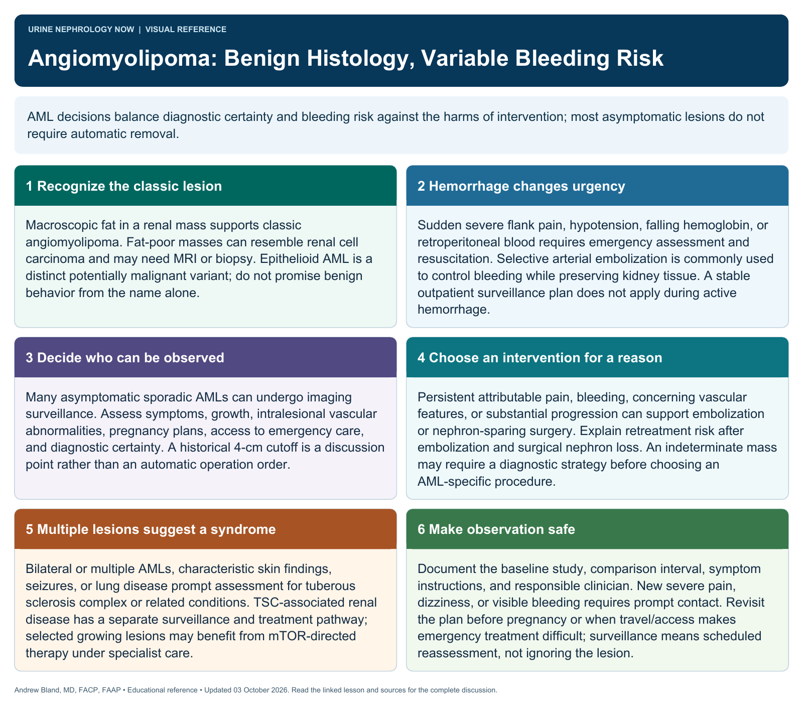
Angiomyolipoma management balances bleeding risk and diagnostic confidence with the value of preserving renal tissue.
Text version
A vascular, muscle, and fat-containing lesion
Renal angiomyolipoma is commonly benign and may occur sporadically or with tuberous sclerosis complex. Fat-rich lesions can have recognizable imaging features, while fat-poor or epithelioid variants require more careful assessment.
Recognize a bleeding emergency
Acute flank or abdominal pain, hypotension, anemia, or evidence of retroperitoneal hemorrhage requires urgent evaluation. A known renal lesion does not make every new pain episode benign or suitable for routine surveillance.
Assess lesion and patient factors
Review imaging characteristics, size and growth, intralesional vascular features, symptoms, reproductive context, and associated syndromic disease. Consider the possibility of a different tumor when imaging is atypical or the diagnosis remains uncertain.
Choose observation or intervention
Surveillance is suitable for many asymptomatic lesions. Selected patients may need embolization, nephron-sparing surgery, or another approach based on bleeding risk, symptoms, anatomy, and the likelihood of preserving kidney function.
Recognize syndromic treatment options
In tuberous sclerosis-associated disease, systemic pathway-directed therapy may be relevant under specialist care. Its indications, monitoring, and adverse effects differ from the management of a small sporadic asymptomatic angiomyolipoma.
Do not use size alone
A historical size threshold should not automatically trigger intervention in every patient. Explain the uncertainty, agree on follow-up ownership, and identify the symptoms or imaging changes that would alter the management plan.
Continue learning
This graphic summarizes a topic. The full educational pages provide the broader discussion and references.