Visual summary
First establish the correct imaging category; then choose surveillance or intervention using tumor features and the person’s competing risks.
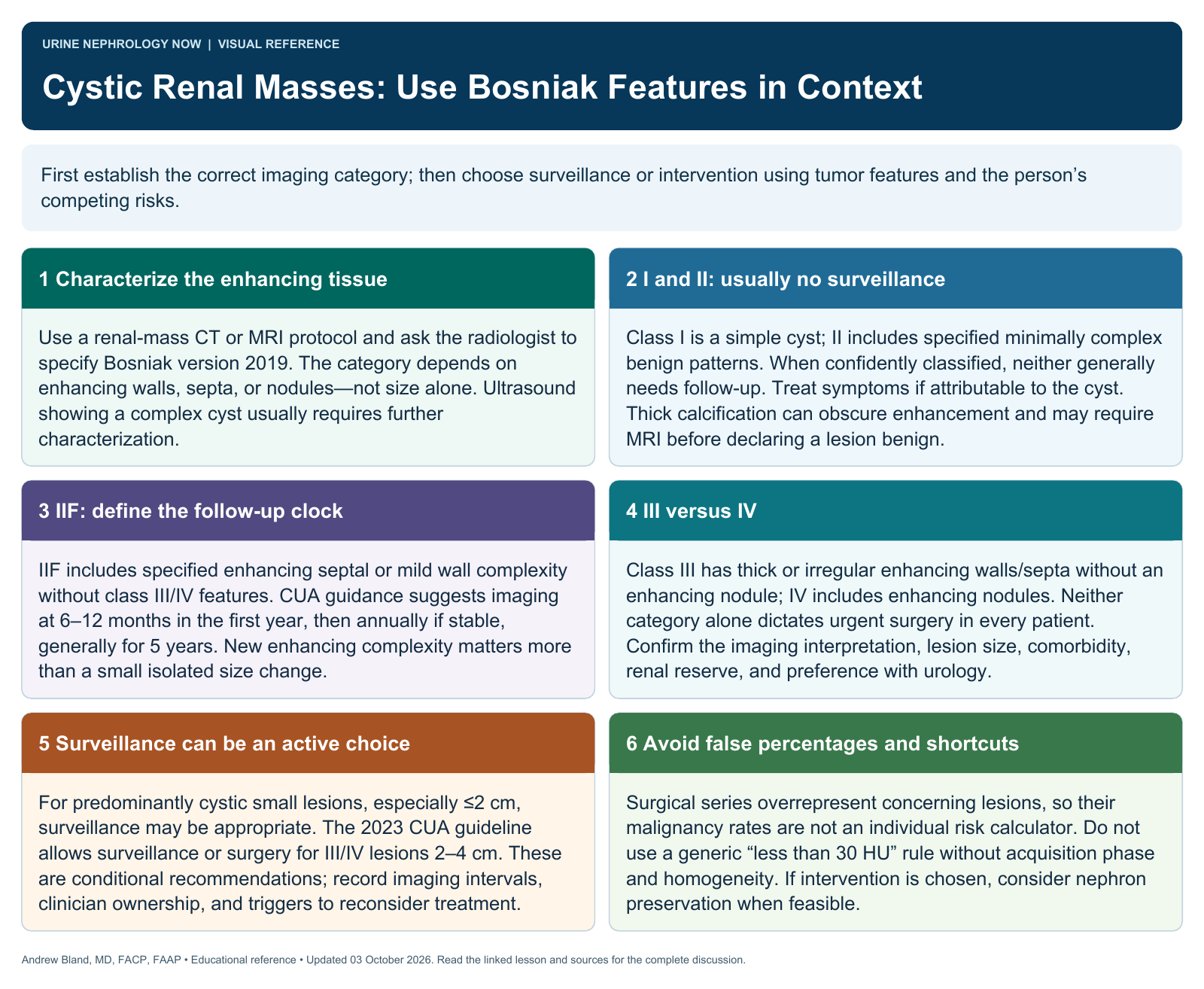
Text version
Characterize the enhancing tissue
Use a renal-mass CT or MRI protocol and ask the radiologist to specify Bosniak version 2019. The category depends on enhancing walls, septa, or nodules—not size alone. Ultrasound showing a complex cyst usually requires further characterization.
I and II: usually no surveillance
Class I is a simple cyst; II includes specified minimally complex benign patterns. When confidently classified, neither generally needs follow-up. Treat symptoms if attributable to the cyst. Thick calcification can obscure enhancement and may require MRI before declaring a lesion benign.
IIF: define the follow-up clock
IIF includes specified enhancing septal or mild wall complexity without class III/IV features. CUA guidance suggests imaging at 6–12 months in the first year, then annually if stable, generally for 5 years. New enhancing complexity matters more than a small isolated size change.
III versus IV
Class III has thick or irregular enhancing walls/septa without an enhancing nodule; IV includes enhancing nodules. Neither category alone dictates urgent surgery in every patient. Confirm the imaging interpretation, lesion size, comorbidity, renal reserve, and preference with urology.
Surveillance can be an active choice
For predominantly cystic small lesions, especially ≤2 cm, surveillance may be appropriate. The 2023 CUA guideline allows surveillance or surgery for III/IV lesions 2–4 cm. These are conditional recommendations; record imaging intervals, clinician ownership, and triggers to reconsider treatment.
Avoid false percentages and shortcuts
Surgical series overrepresent concerning lesions, so their malignancy rates are not an individual risk calculator. Do not use a generic “less than 30 HU” rule without acquisition phase and homogeneity. If intervention is chosen, consider nephron preservation when feasible.