Visual summary
Use sediment and systemic findings to choose the next targeted test; do not diagnose a kidney compartment from a chemistry pattern alone.
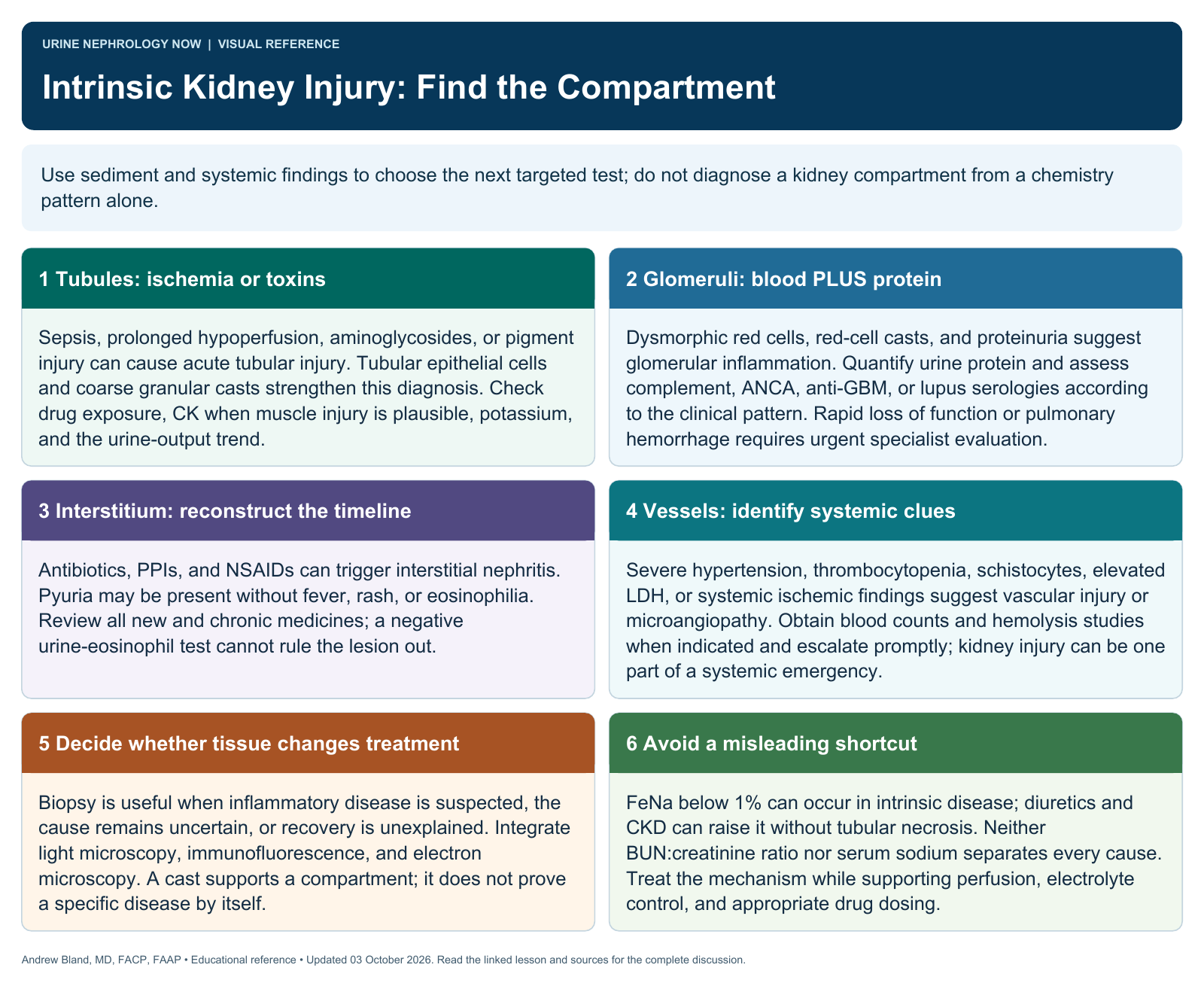
Text version
Tubules: ischemia or toxins
Sepsis, prolonged hypoperfusion, aminoglycosides, or pigment injury can cause acute tubular injury. Tubular epithelial cells and coarse granular casts strengthen this diagnosis. Check drug exposure, CK when muscle injury is plausible, potassium, and the urine-output trend.
Glomeruli: blood PLUS protein
Dysmorphic red cells, red-cell casts, and proteinuria suggest glomerular inflammation. Quantify urine protein and assess complement, ANCA, anti-GBM, or lupus serologies according to the clinical pattern. Rapid loss of function or pulmonary hemorrhage requires urgent specialist evaluation.
Interstitium: reconstruct the timeline
Antibiotics, PPIs, and NSAIDs can trigger interstitial nephritis. Pyuria may be present without fever, rash, or eosinophilia. Review all new and chronic medicines; a negative urine-eosinophil test cannot rule the lesion out.
Vessels: identify systemic clues
Severe hypertension, thrombocytopenia, schistocytes, elevated LDH, or systemic ischemic findings suggest vascular injury or microangiopathy. Obtain blood counts and hemolysis studies when indicated and escalate promptly; kidney injury can be one part of a systemic emergency.
Decide whether tissue changes treatment
Biopsy is useful when inflammatory disease is suspected, the cause remains uncertain, or recovery is unexplained. Integrate light microscopy, immunofluorescence, and electron microscopy. A cast supports a compartment; it does not prove a specific disease by itself.
Avoid a misleading shortcut
FeNa below 1% can occur in intrinsic disease; diuretics and CKD can raise it without tubular necrosis. Neither BUN:creatinine ratio nor serum sodium separates every cause. Treat the mechanism while supporting perfusion, electrolyte control, and appropriate drug dosing.