Visual summary
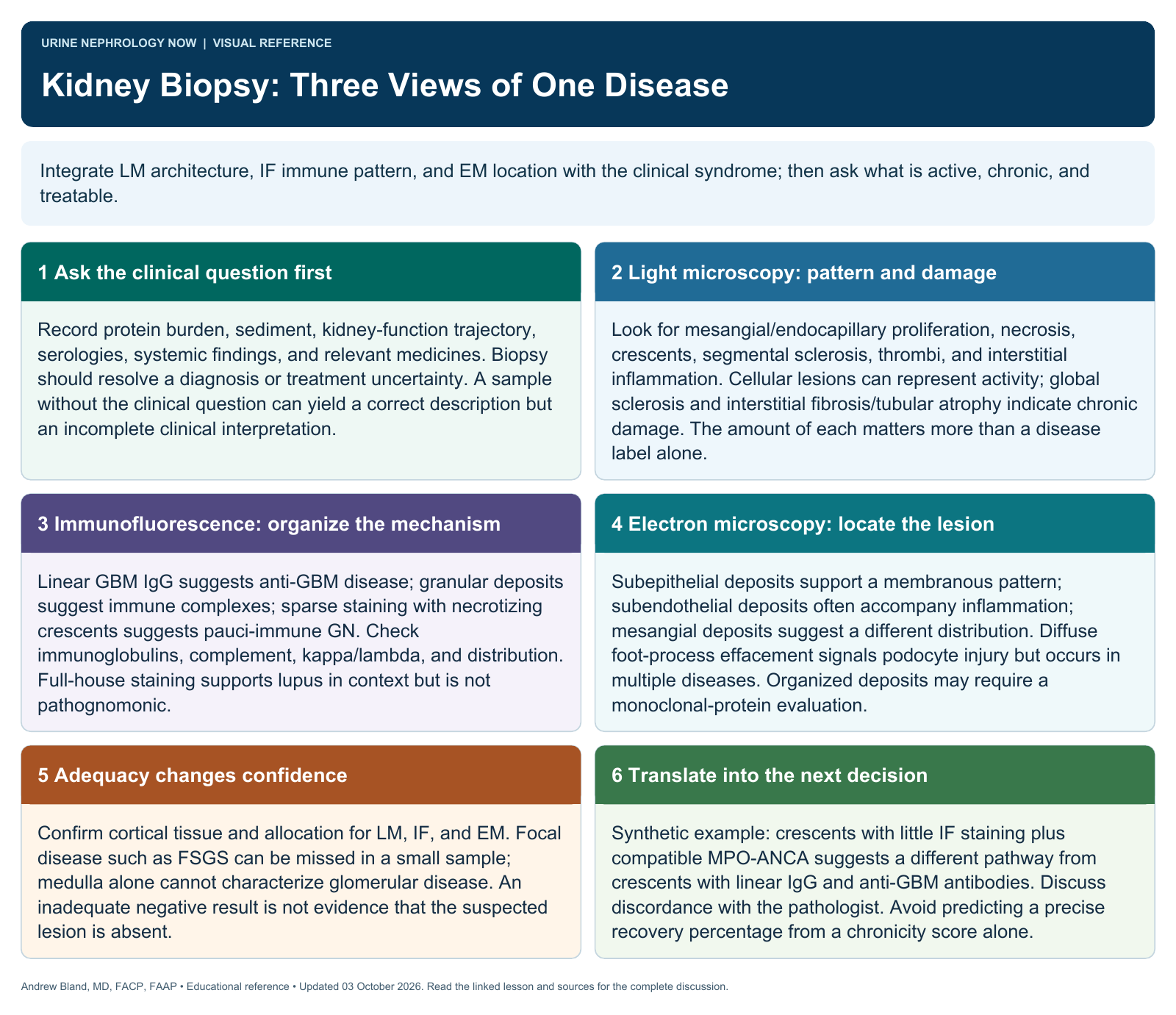
A biopsy is an integrated explanation of injury; specimen quality and clinical correlation are as important as the pattern name.
Text version
Architecture, immune signal, ultrastructure
Light microscopy shows the injury pattern, immunofluorescence identifies immune staining, and electron microscopy locates deposits and structural abnormalities. The strongest interpretation comes from integrating all three with the clinical presentation.
Recognize activity versus chronicity
Cellular inflammation, necrosis, and some crescents can indicate active injury; sclerosis and interstitial fibrosis signal chronic damage. These findings influence prognosis, but no single percentage should determine treatment without context.
Assess sample adequacy
Confirm that the tissue contains relevant compartments and has been allocated correctly for the required studies. Focal disease can be missed by sampling, while insufficient tissue limits confidence even when the report offers a favored diagnosis.
Read the staining pattern
Linear, granular, and pauci-immune patterns narrow the differential but are not free-standing clinical diagnoses. The immunoglobulin, complement, light-chain distribution, intensity, and location help distinguish overlapping entities.
Connect deposits and structure
Subendothelial, mesangial, and subepithelial deposits suggest different mechanisms. Foot-process effacement indicates podocyte injury but occurs in more than one disease. Correlate ultrastructure with proteinuria, hematuria, and serologic findings.
Translate the report into action
Ask what is active, what is chronic, what caused it, and which uncertainty remains. Discuss discordant clinical and pathologic findings with the renal pathologist, and avoid promising reversibility from a classification label alone.
Continue learning
This graphic summarizes a topic. The full educational pages provide the broader discussion and references.