Visual summary
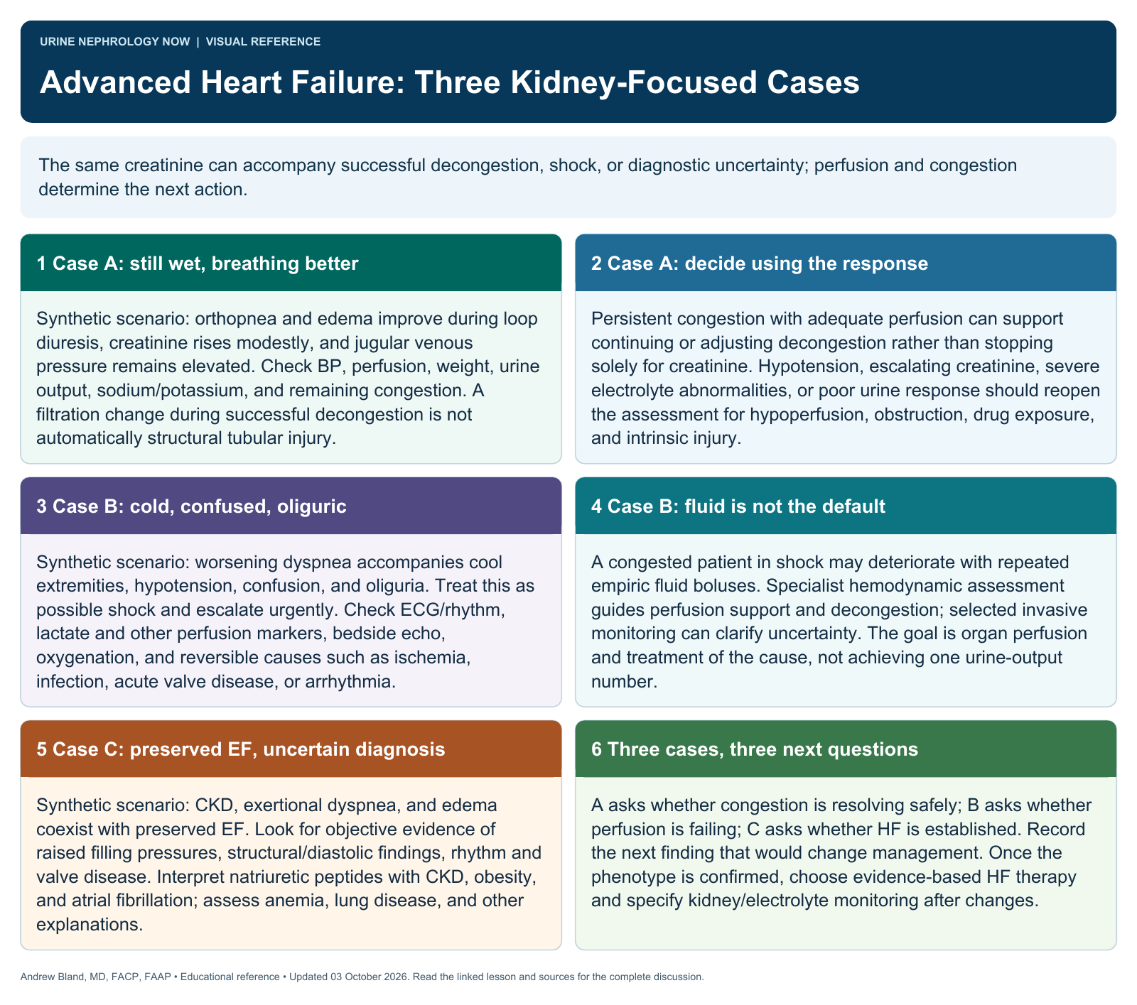
The same creatinine can accompany successful decongestion, shock, or diagnostic uncertainty; perfusion and congestion determine the next action.
Text version
Case A: still wet, breathing better
Synthetic scenario: orthopnea and edema improve during loop diuresis, creatinine rises modestly, and jugular venous pressure remains elevated. Check BP, perfusion, weight, urine output, sodium/potassium, and remaining congestion. A filtration change during successful decongestion is not automatically structural tubular injury.
Case A: decide using the response
Persistent congestion with adequate perfusion can support continuing or adjusting decongestion rather than stopping solely for creatinine. Hypotension, escalating creatinine, severe electrolyte abnormalities, or poor urine response should reopen the assessment for hypoperfusion, obstruction, drug exposure, and intrinsic injury.
Case B: cold, confused, oliguric
Synthetic scenario: worsening dyspnea accompanies cool extremities, hypotension, confusion, and oliguria. Treat this as possible shock and escalate urgently. Check ECG/rhythm, lactate and other perfusion markers, bedside echo, oxygenation, and reversible causes such as ischemia, infection, acute valve disease, or arrhythmia.
Case B: fluid is not the default
A congested patient in shock may deteriorate with repeated empiric fluid boluses. Specialist hemodynamic assessment guides perfusion support and decongestion; selected invasive monitoring can clarify uncertainty. The goal is organ perfusion and treatment of the cause, not achieving one urine-output number.
Case C: preserved EF, uncertain diagnosis
Synthetic scenario: CKD, exertional dyspnea, and edema coexist with preserved EF. Look for objective evidence of raised filling pressures, structural/diastolic findings, rhythm and valve disease. Interpret natriuretic peptides with CKD, obesity, and atrial fibrillation; assess anemia, lung disease, and other explanations.
Three cases, three next questions
A asks whether congestion is resolving safely; B asks whether perfusion is failing; C asks whether HF is established. Record the next finding that would change management. Once the phenotype is confirmed, choose evidence-based HF therapy and specify kidney/electrolyte monitoring after changes.