Visual summary
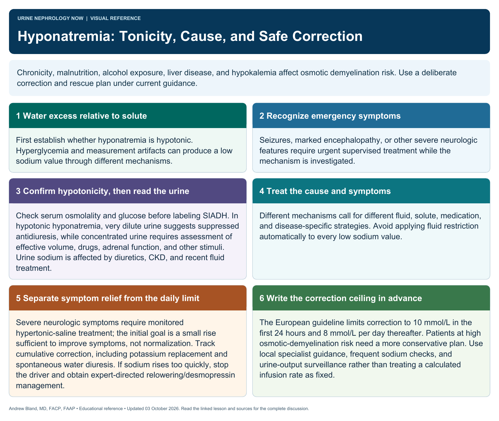
Chronicity, malnutrition, alcohol exposure, liver disease, and hypokalemia affect osmotic demyelination risk. Use a deliberate correction and rescue plan under current guidance.
Text version
Water excess relative to solute
First establish whether hyponatremia is hypotonic. Hyperglycemia and measurement artifacts can produce a low sodium value through different mechanisms.
Recognize emergency symptoms
Seizures, marked encephalopathy, or other severe neurologic features require urgent supervised treatment while the mechanism is investigated.
Confirm hypotonicity, then read the urine
Check serum osmolality and glucose before labeling SIADH. In hypotonic hyponatremia, very dilute urine suggests suppressed antidiuresis, while concentrated urine requires assessment of effective volume, drugs, adrenal function, and other stimuli. Urine sodium is affected by diuretics, CKD, and recent fluid treatment.
Treat the cause and symptoms
Different mechanisms call for different fluid, solute, medication, and disease-specific strategies. Avoid applying fluid restriction automatically to every low sodium value.
Separate symptom relief from the daily limit
Severe neurologic symptoms require monitored hypertonic-saline treatment; the initial goal is a small rise sufficient to improve symptoms, not normalization. Track cumulative correction, including potassium replacement and spontaneous water diuresis. If sodium rises too quickly, stop the driver and obtain expert-directed relowering/desmopressin management.
Write the correction ceiling in advance
The European guideline limits correction to 10 mmol/L in the first 24 hours and 8 mmol/L per day thereafter. Patients at high osmotic-demyelination risk need a more conservative plan. Use local specialist guidance, frequent sodium checks, and urine-output surveillance rather than treating a calculated infusion rate as fixed.