Visual summary
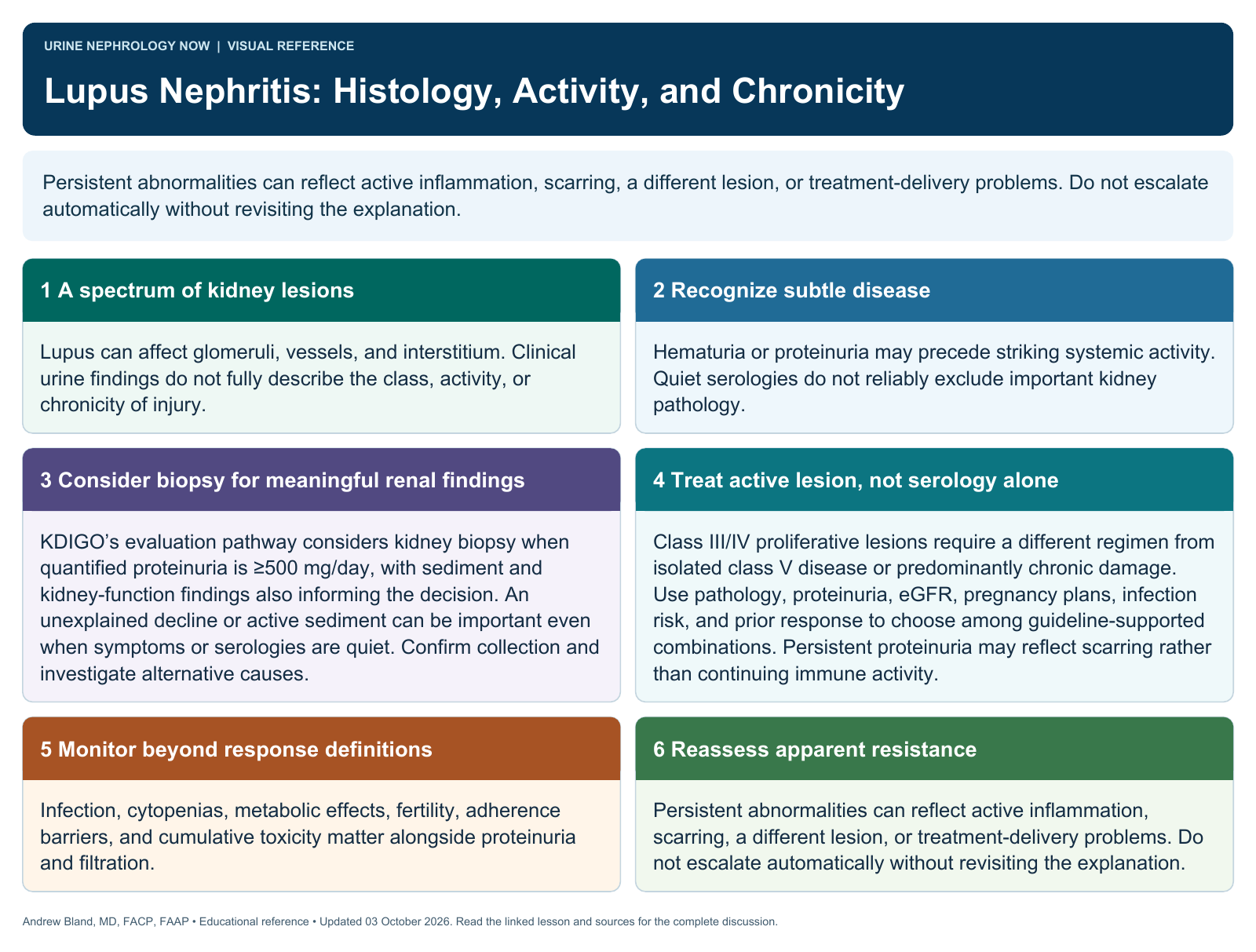
Persistent abnormalities can reflect active inflammation, scarring, a different lesion, or treatment-delivery problems. Do not escalate automatically without revisiting the explanation.
Text version
A spectrum of kidney lesions
Lupus can affect glomeruli, vessels, and interstitium. Clinical urine findings do not fully describe the class, activity, or chronicity of injury.
Recognize subtle disease
Hematuria or proteinuria may precede striking systemic activity. Quiet serologies do not reliably exclude important kidney pathology.
Consider biopsy for meaningful renal findings
KDIGO’s evaluation pathway considers kidney biopsy when quantified proteinuria is ≥500 mg/day, with sediment and kidney-function findings also informing the decision. An unexplained decline or active sediment can be important even when symptoms or serologies are quiet. Confirm collection and investigate alternative causes.
Treat active lesion, not serology alone
Class III/IV proliferative lesions require a different regimen from isolated class V disease or predominantly chronic damage. Use pathology, proteinuria, eGFR, pregnancy plans, infection risk, and prior response to choose among guideline-supported combinations. Persistent proteinuria may reflect scarring rather than continuing immune activity.
Monitor beyond response definitions
Infection, cytopenias, metabolic effects, fertility, adherence barriers, and cumulative toxicity matter alongside proteinuria and filtration.
Reassess apparent resistance
Persistent abnormalities can reflect active inflammation, scarring, a different lesion, or treatment-delivery problems. Do not escalate automatically without revisiting the explanation.