Visual summary
Failure to respond as expected should prompt review of diagnosis, adherence, secondary causes, and whether additional pathology is present.
Text version
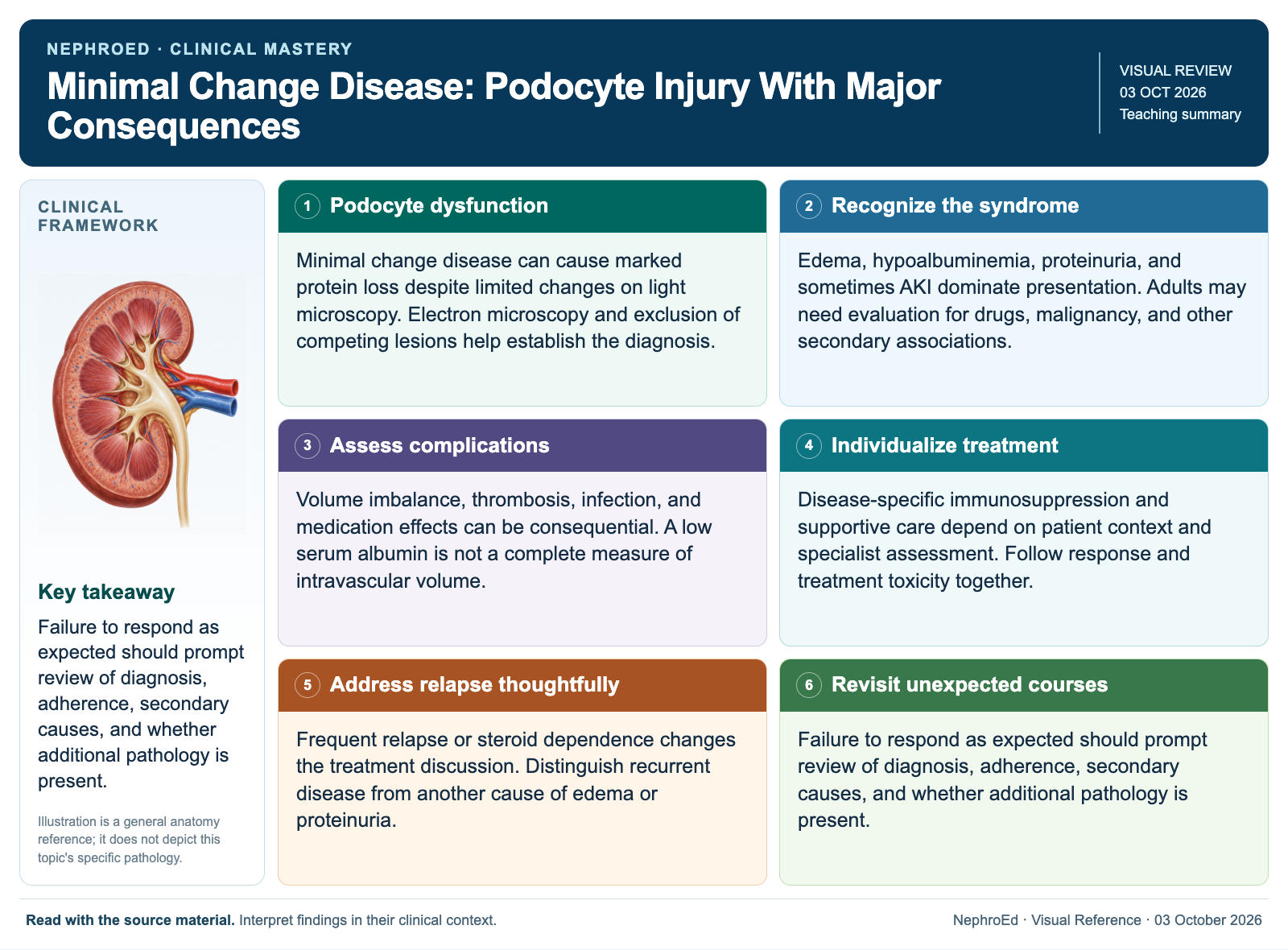
Podocyte dysfunction
Minimal change disease can cause marked protein loss despite limited changes on light microscopy. Electron microscopy and exclusion of competing lesions help establish the diagnosis.
Recognize the syndrome
Edema, hypoalbuminemia, proteinuria, and sometimes AKI dominate presentation. Adults may need evaluation for drugs, malignancy, and other secondary associations.
Assess complications
Volume imbalance, thrombosis, infection, and medication effects can be consequential. A low serum albumin is not a complete measure of intravascular volume.
Individualize treatment
Disease-specific immunosuppression and supportive care depend on patient context and specialist assessment. Follow response and treatment toxicity together.
Address relapse thoughtfully
Frequent relapse or steroid dependence changes the treatment discussion. Distinguish recurrent disease from another cause of edema or proteinuria.
Revisit unexpected courses
Failure to respond as expected should prompt review of diagnosis, adherence, secondary causes, and whether additional pathology is present.
Continue learning
This graphic summarizes a topic. The full educational pages provide the broader discussion and references.