Visual summary
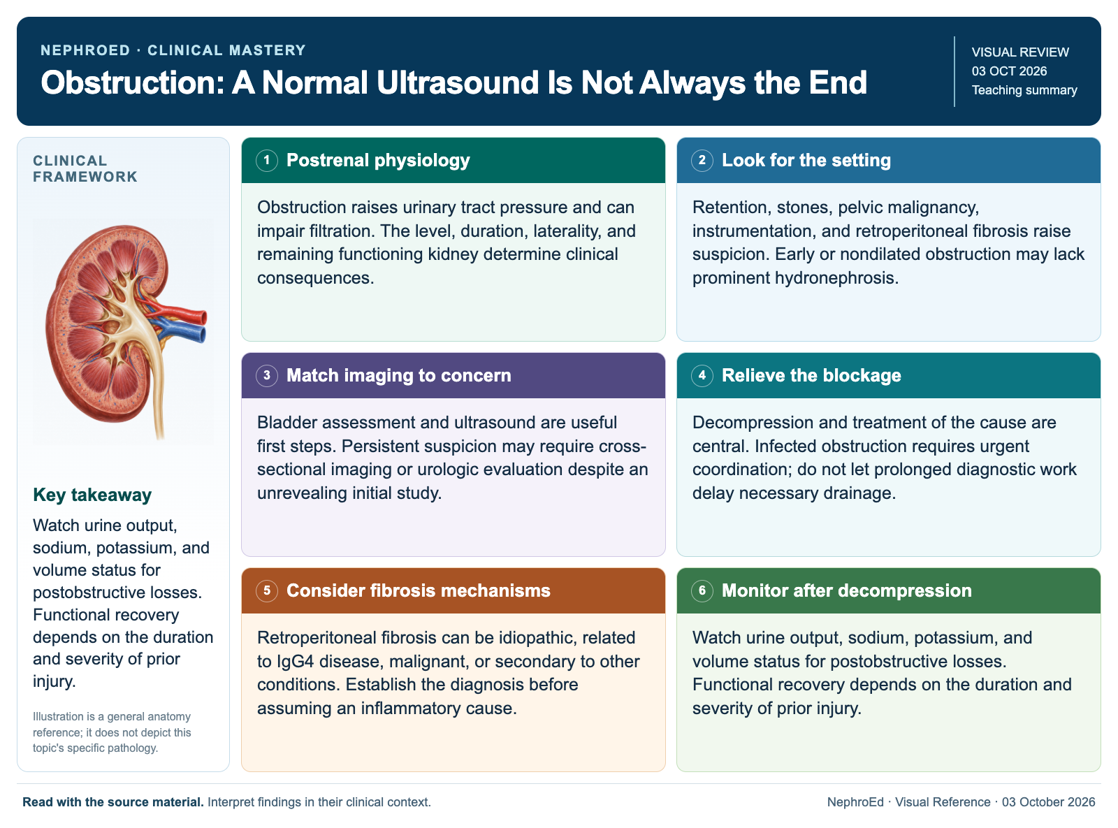
Watch urine output, sodium, potassium, and volume status for postobstructive losses. Functional recovery depends on the duration and severity of prior injury.
Text version
Postrenal physiology
Obstruction raises urinary tract pressure and can impair filtration. The level, duration, laterality, and remaining functioning kidney determine clinical consequences.
Look for the setting
Retention, stones, pelvic malignancy, instrumentation, and retroperitoneal fibrosis raise suspicion. Early or nondilated obstruction may lack prominent hydronephrosis.
Match imaging to concern
Bladder assessment and ultrasound are useful first steps. Persistent suspicion may require cross-sectional imaging or urologic evaluation despite an unrevealing initial study.
Relieve the blockage
Decompression and treatment of the cause are central. Infected obstruction requires urgent coordination; do not let prolonged diagnostic work delay necessary drainage.
Consider fibrosis mechanisms
Retroperitoneal fibrosis can be idiopathic, related to IgG4 disease, malignant, or secondary to other conditions. Establish the diagnosis before assuming an inflammatory cause.
Monitor after decompression
Watch urine output, sodium, potassium, and volume status for postobstructive losses. Functional recovery depends on the duration and severity of prior injury.
Continue learning
This graphic summarizes a topic. The full educational pages provide the broader discussion and references.