Visual summary
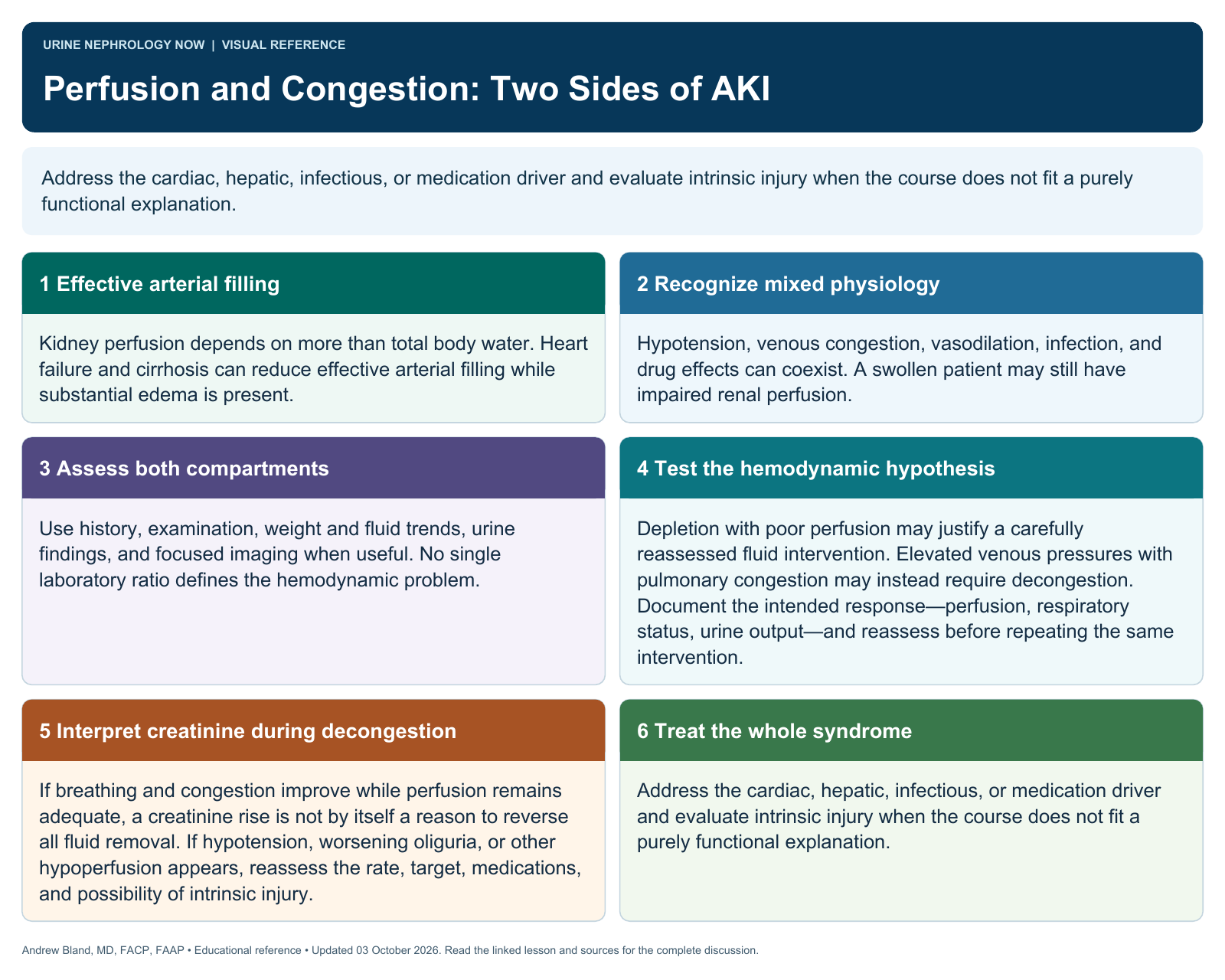
Address the cardiac, hepatic, infectious, or medication driver and evaluate intrinsic injury when the course does not fit a purely functional explanation.
Text version
Effective arterial filling
Kidney perfusion depends on more than total body water. Heart failure and cirrhosis can reduce effective arterial filling while substantial edema is present.
Recognize mixed physiology
Hypotension, venous congestion, vasodilation, infection, and drug effects can coexist. A swollen patient may still have impaired renal perfusion.
Assess both compartments
Use history, examination, weight and fluid trends, urine findings, and focused imaging when useful. No single laboratory ratio defines the hemodynamic problem.
Test the hemodynamic hypothesis
Depletion with poor perfusion may justify a carefully reassessed fluid intervention. Elevated venous pressures with pulmonary congestion may instead require decongestion. Document the intended response—perfusion, respiratory status, urine output—and reassess before repeating the same intervention.
Interpret creatinine during decongestion
If breathing and congestion improve while perfusion remains adequate, a creatinine rise is not by itself a reason to reverse all fluid removal. If hypotension, worsening oliguria, or other hypoperfusion appears, reassess the rate, target, medications, and possibility of intrinsic injury.
Treat the whole syndrome
Address the cardiac, hepatic, infectious, or medication driver and evaluate intrinsic injury when the course does not fit a purely functional explanation.