Visual summary
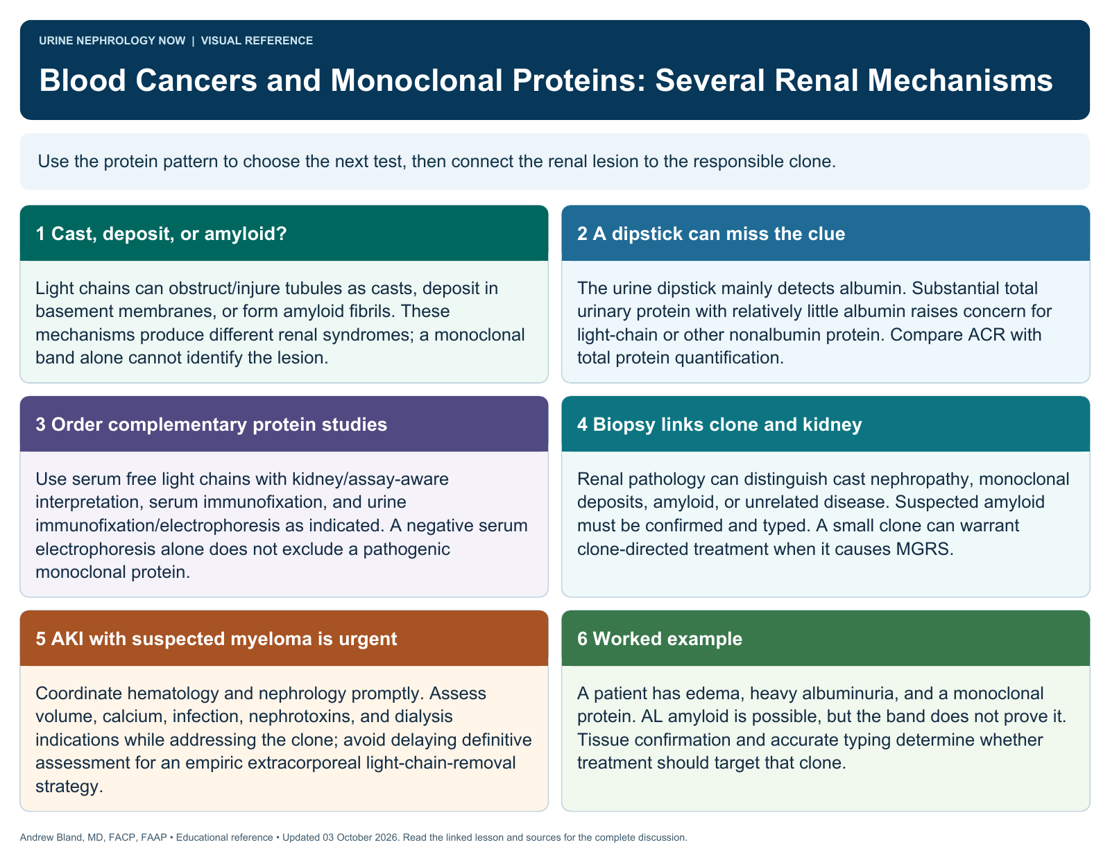
Use the protein pattern to choose the next test, then connect the renal lesion to the responsible clone.
Text version
Cast, deposit, or amyloid?
Light chains can obstruct/injure tubules as casts, deposit in basement membranes, or form amyloid fibrils. These mechanisms produce different renal syndromes; a monoclonal band alone cannot identify the lesion.
A dipstick can miss the clue
The urine dipstick mainly detects albumin. Substantial total urinary protein with relatively little albumin raises concern for light-chain or other nonalbumin protein. Compare ACR with total protein quantification.
Order complementary protein studies
Use serum free light chains with kidney/assay-aware interpretation, serum immunofixation, and urine immunofixation/electrophoresis as indicated. A negative serum electrophoresis alone does not exclude a pathogenic monoclonal protein.
Biopsy links clone and kidney
Renal pathology can distinguish cast nephropathy, monoclonal deposits, amyloid, or unrelated disease. Suspected amyloid must be confirmed and typed. A small clone can warrant clone-directed treatment when it causes MGRS.
AKI with suspected myeloma is urgent
Coordinate hematology and nephrology promptly. Assess volume, calcium, infection, nephrotoxins, and dialysis indications while addressing the clone; avoid delaying definitive assessment for an empiric extracorporeal light-chain-removal strategy.
Worked example
A patient has edema, heavy albuminuria, and a monoclonal protein. AL amyloid is possible, but the band does not prove it. Tissue confirmation and accurate typing determine whether treatment should target that clone.