Visual summary
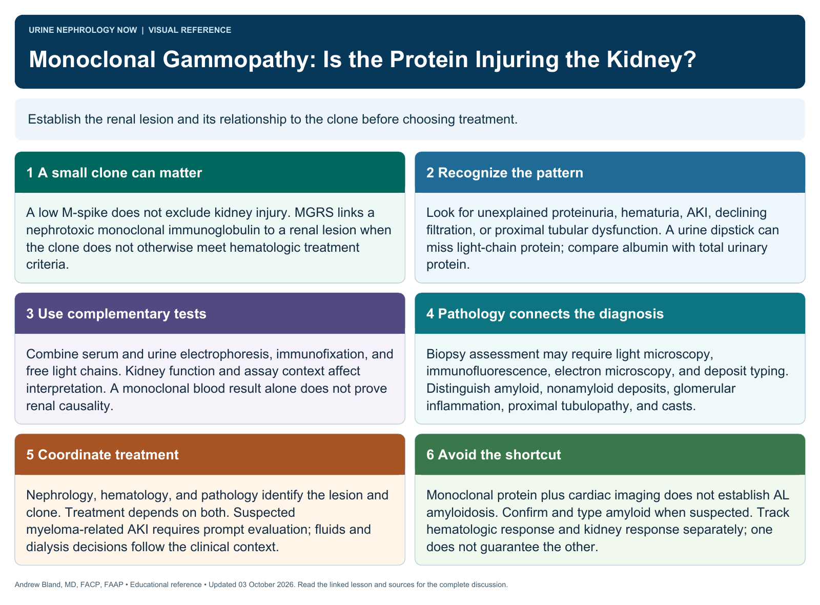
Establish the renal lesion and its relationship to the clone before choosing treatment.
Text version
A small clone can matter
A low M-spike does not exclude kidney injury. MGRS links a nephrotoxic monoclonal immunoglobulin to a renal lesion when the clone does not otherwise meet hematologic treatment criteria.
Recognize the pattern
Look for unexplained proteinuria, hematuria, AKI, declining filtration, or proximal tubular dysfunction. A urine dipstick can miss light-chain protein; compare albumin with total urinary protein.
Use complementary tests
Combine serum and urine electrophoresis, immunofixation, and free light chains. Kidney function and assay context affect interpretation. A monoclonal blood result alone does not prove renal causality.
Pathology connects the diagnosis
Biopsy assessment may require light microscopy, immunofluorescence, electron microscopy, and deposit typing. Distinguish amyloid, nonamyloid deposits, glomerular inflammation, proximal tubulopathy, and casts.
Coordinate treatment
Nephrology, hematology, and pathology identify the lesion and clone. Treatment depends on both. Suspected myeloma-related AKI requires prompt evaluation; fluids and dialysis decisions follow the clinical context.
Avoid the shortcut
Monoclonal protein plus cardiac imaging does not establish AL amyloidosis. Confirm and type amyloid when suspected. Track hematologic response and kidney response separately; one does not guarantee the other.
Continue learning
This graphic summarizes a topic. The full educational pages provide the broader discussion and references.