Visual summary
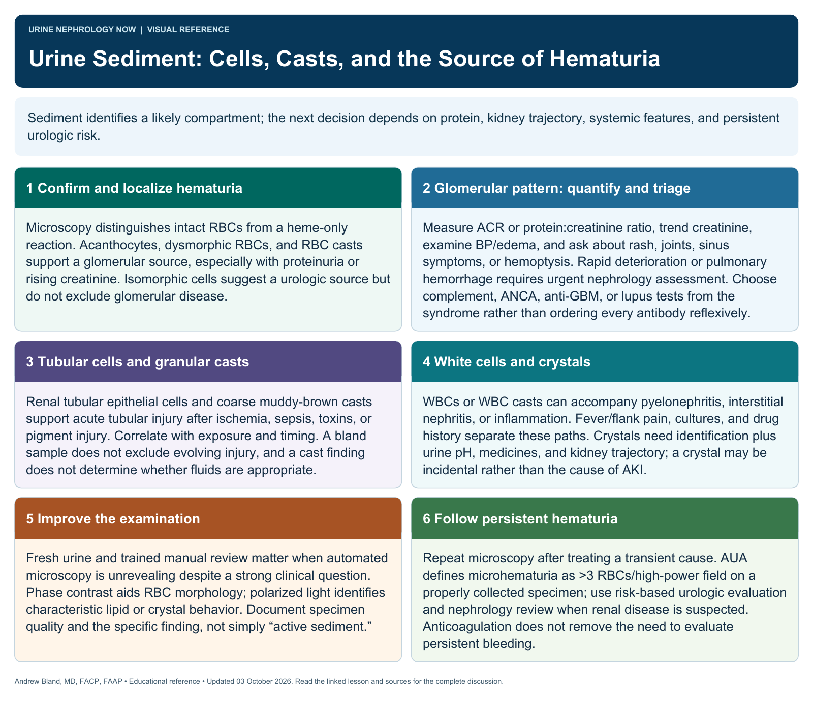
Sediment identifies a likely compartment; the next decision depends on protein, kidney trajectory, systemic features, and persistent urologic risk.
Text version
Confirm and localize hematuria
Microscopy distinguishes intact RBCs from a heme-only reaction. Acanthocytes, dysmorphic RBCs, and RBC casts support a glomerular source, especially with proteinuria or rising creatinine. Isomorphic cells suggest a urologic source but do not exclude glomerular disease.
Glomerular pattern: quantify and triage
Measure ACR or protein:creatinine ratio, trend creatinine, examine BP/edema, and ask about rash, joints, sinus symptoms, or hemoptysis. Rapid deterioration or pulmonary hemorrhage requires urgent nephrology assessment. Choose complement, ANCA, anti-GBM, or lupus tests from the syndrome rather than ordering every antibody reflexively.
Tubular cells and granular casts
Renal tubular epithelial cells and coarse muddy-brown casts support acute tubular injury after ischemia, sepsis, toxins, or pigment injury. Correlate with exposure and timing. A bland sample does not exclude evolving injury, and a cast finding does not determine whether fluids are appropriate.
White cells and crystals
WBCs or WBC casts can accompany pyelonephritis, interstitial nephritis, or inflammation. Fever/flank pain, cultures, and drug history separate these paths. Crystals need identification plus urine pH, medicines, and kidney trajectory; a crystal may be incidental rather than the cause of AKI.
Improve the examination
Fresh urine and trained manual review matter when automated microscopy is unrevealing despite a strong clinical question. Phase contrast aids RBC morphology; polarized light identifies characteristic lipid or crystal behavior. Document specimen quality and the specific finding, not simply “active sediment.”
Follow persistent hematuria
Repeat microscopy after treating a transient cause. AUA defines microhematuria as >3 RBCs/high-power field on a properly collected specimen; use risk-based urologic evaluation and nephrology review when renal disease is suspected. Anticoagulation does not remove the need to evaluate persistent bleeding.