Visual summary
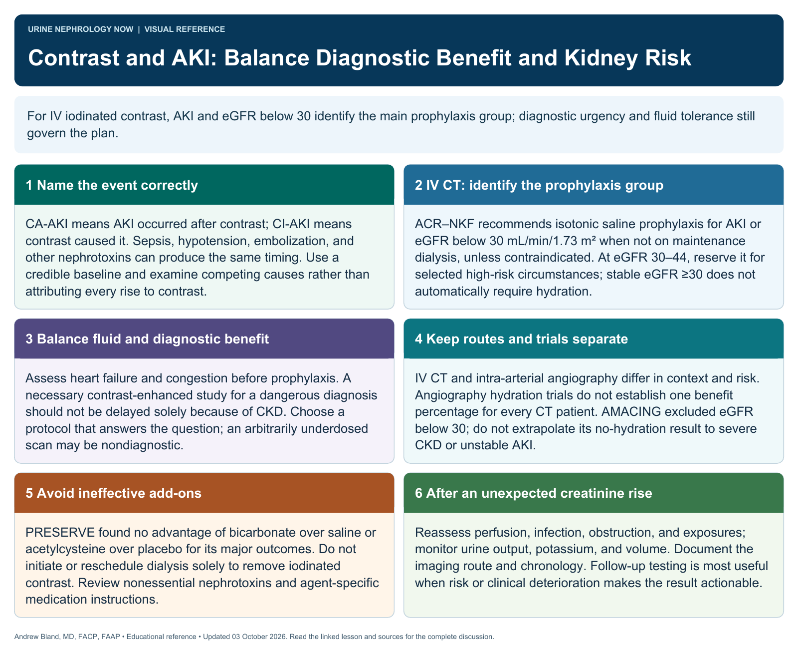
For IV iodinated contrast, AKI and eGFR below 30 identify the main prophylaxis group; diagnostic urgency and fluid tolerance still govern the plan.
Text version
Name the event correctly
CA-AKI means AKI occurred after contrast; CI-AKI means contrast caused it. Sepsis, hypotension, embolization, and other nephrotoxins can produce the same timing. Use a credible baseline and examine competing causes rather than attributing every rise to contrast.
IV CT: identify the prophylaxis group
ACR–NKF recommends isotonic saline prophylaxis for AKI or eGFR below 30 mL/min/1.73 m² when not on maintenance dialysis, unless contraindicated. At eGFR 30–44, reserve it for selected high-risk circumstances; stable eGFR ≥30 does not automatically require hydration.
Balance fluid and diagnostic benefit
Assess heart failure and congestion before prophylaxis. A necessary contrast-enhanced study for a dangerous diagnosis should not be delayed solely because of CKD. Choose a protocol that answers the question; an arbitrarily underdosed scan may be nondiagnostic.
Keep routes and trials separate
IV CT and intra-arterial angiography differ in context and risk. Angiography hydration trials do not establish one benefit percentage for every CT patient. AMACING excluded eGFR below 30; do not extrapolate its no-hydration result to severe CKD or unstable AKI.
Avoid ineffective add-ons
PRESERVE found no advantage of bicarbonate over saline or acetylcysteine over placebo for its major outcomes. Do not initiate or reschedule dialysis solely to remove iodinated contrast. Review nonessential nephrotoxins and agent-specific medication instructions.
After an unexpected creatinine rise
Reassess perfusion, infection, obstruction, and exposures; monitor urine output, potassium, and volume. Document the imaging route and chronology. Follow-up testing is most useful when risk or clinical deterioration makes the result actionable.