Visual summary
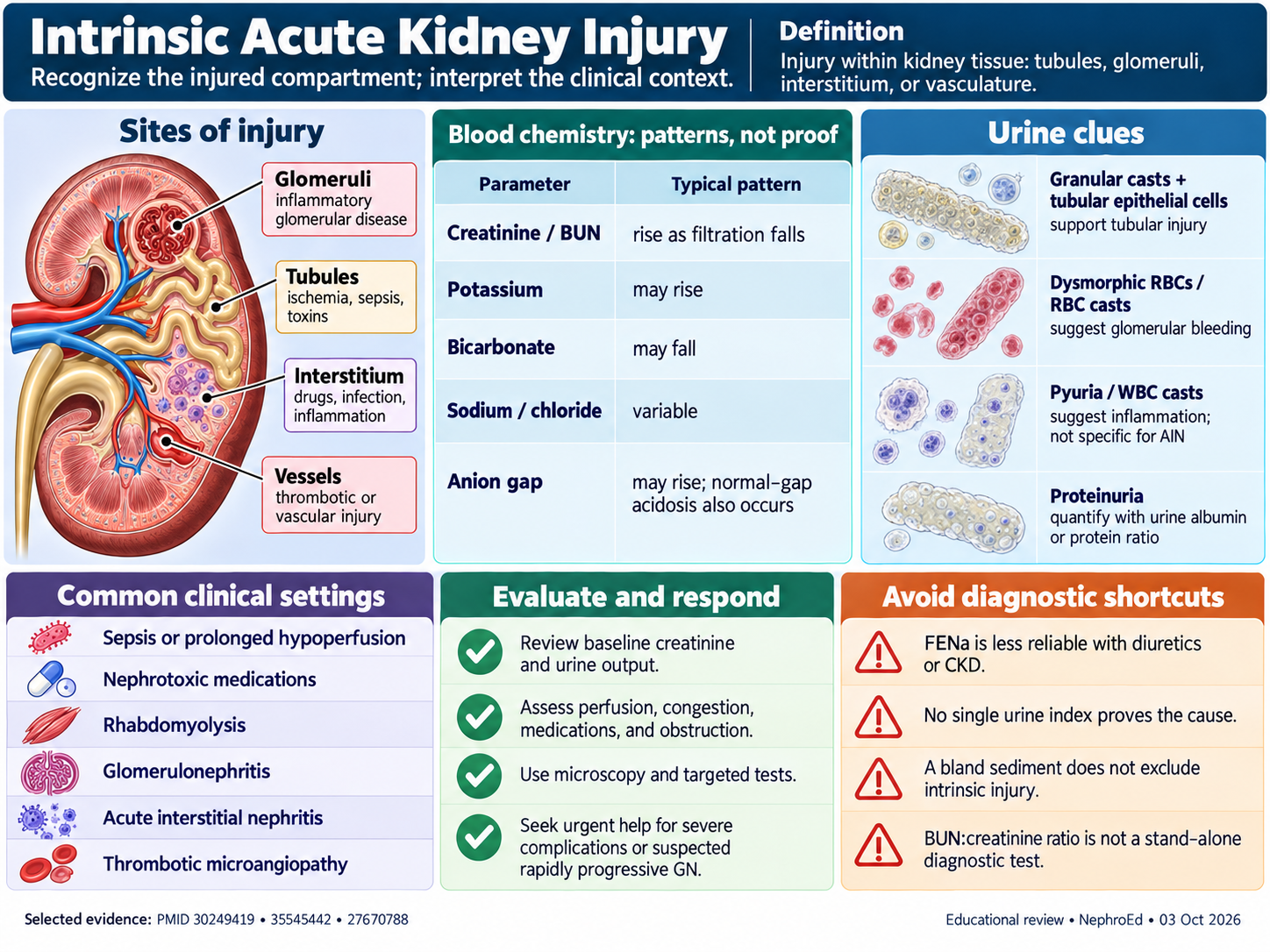
Recognize the injured compartment and interpret the clinical context.
Text version
Sites of injury
Glomeruli: inflammatory glomerular disease. Tubules: ischemia, sepsis, toxins. Interstitium: drugs, infection, inflammation. Vessels: thrombotic or vascular injury.
Blood chemistry: patterns, not proof
Creatinine and BUN rise as filtration falls. Potassium may rise and bicarbonate may fall. Sodium and chloride are variable. The anion gap may rise; normal-gap acidosis also occurs.
Urine clues
Granular casts and tubular epithelial cells support tubular injury. Dysmorphic red cells or red cell casts suggest glomerular bleeding. Pyuria or white cell casts suggest inflammation but are not specific for acute interstitial nephritis. Quantify proteinuria with a urine albumin or protein ratio.
Common clinical settings
Sepsis or prolonged hypoperfusion, nephrotoxic medications, rhabdomyolysis, glomerulonephritis, acute interstitial nephritis, and thrombotic microangiopathy.
Evaluate and respond
Review baseline creatinine and urine output. Assess perfusion, congestion, medications, and obstruction. Use microscopy and targeted tests. Seek urgent help for severe complications or suspected rapidly progressive glomerulonephritis.
Avoid diagnostic shortcuts
FENa is less reliable with diuretics or CKD. No single urine index proves cause. A bland sediment does not exclude intrinsic injury. BUN:creatinine ratio is not a stand-alone diagnostic test.
Continue learning
This graphic summarizes a topic. The full educational pages provide the broader discussion and references.