Visual summary
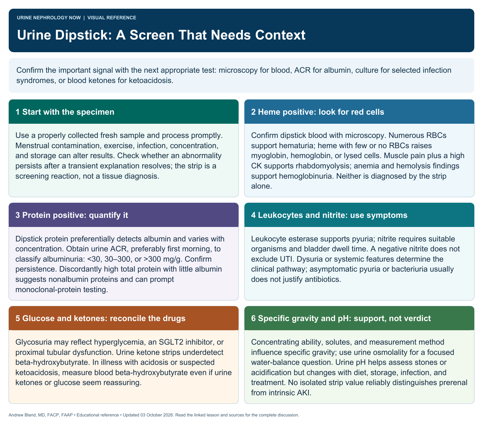
Use dipstick findings to guide the next question, then confirm abnormalities with the test that actually measures the suspected process.
Text version
Chemistry is not microscopy
Dipsticks detect chemical reactions, including heme and selected proteins, rather than directly identifying cells or casts. They are useful screening tools, but results depend on urine concentration, collection, storage, and interfering substances.
Recognize discordant patterns
A blood-positive strip with few red cells can suggest hemoglobin or myoglobin, while false reactions also occur. Protein dipsticks favor albumin and can miss other protein types; a negative strip does not exclude every important renal disorder.
Check specimen quality
Review symptoms, collection method, contamination risk, timing, and whether the sample was processed promptly. Compare specific gravity, pH, and the clinical context before concluding that a weak or negative reaction is reassuring.
Confirm the clinically relevant finding
Use microscopy for cellular and cast assessment and quantitative urine albumin or protein testing when appropriate. Glycosuria may reflect hyperglycemia, medications, or tubular handling; ketone strips do not measure every circulating ketone equally.
Interpret infection markers carefully
Leukocyte esterase and nitrite can support a urinary infection assessment but do not establish infection without symptoms and context. A negative nitrite is not exclusionary, while pyuria or bacteriuria alone does not mandate antibiotics.
Avoid diagnostic shortcuts
A single strip result should not diagnose glomerulonephritis, rhabdomyolysis, or UTI by itself. Investigate persistent abnormalities and reconcile disagreement with microscopy, quantitative tests, medications, and the patient’s trajectory.
Continue learning
This graphic summarizes a topic. The full educational pages provide the broader discussion and references.