Visual reference
Browse the visual reference library · Download lesson Markdown
📚 Why Eating Disorders Matter to the Nephrologist
Eating disorders — particularly anorexia nervosa (AN) and bulimia nervosa (BN) — are psychiatric conditions with profound medical consequences. Nephrologists encounter these patients because of their characteristic electrolyte derangements, acid-base disturbances, and acute kidney injury. The purging behaviors in bulimia and the severe caloric restriction in anorexia each produce distinct metabolic signatures that serve as diagnostic clues.
An estimated 5-10% of the general population has some form of disordered eating, and the renal complications are underrecognized. The nephrologist may be the first to suspect an occult eating disorder based on a puzzling electrolyte pattern.
⚠️ Clinical Significance
- Eating disorders have the highest mortality rate of any psychiatric illness (~5-10% over 10 years)
- Electrolyte abnormalities cause the most acute life-threatening complications (arrhythmia, seizure)
- Patients typically conceal their behaviors — the lab pattern may be the only clue
- Refeeding syndrome can cause fatal hypophosphatemia if nutrition is restarted too aggressively
🔬 Purging Methods & Their Electrolyte Signatures
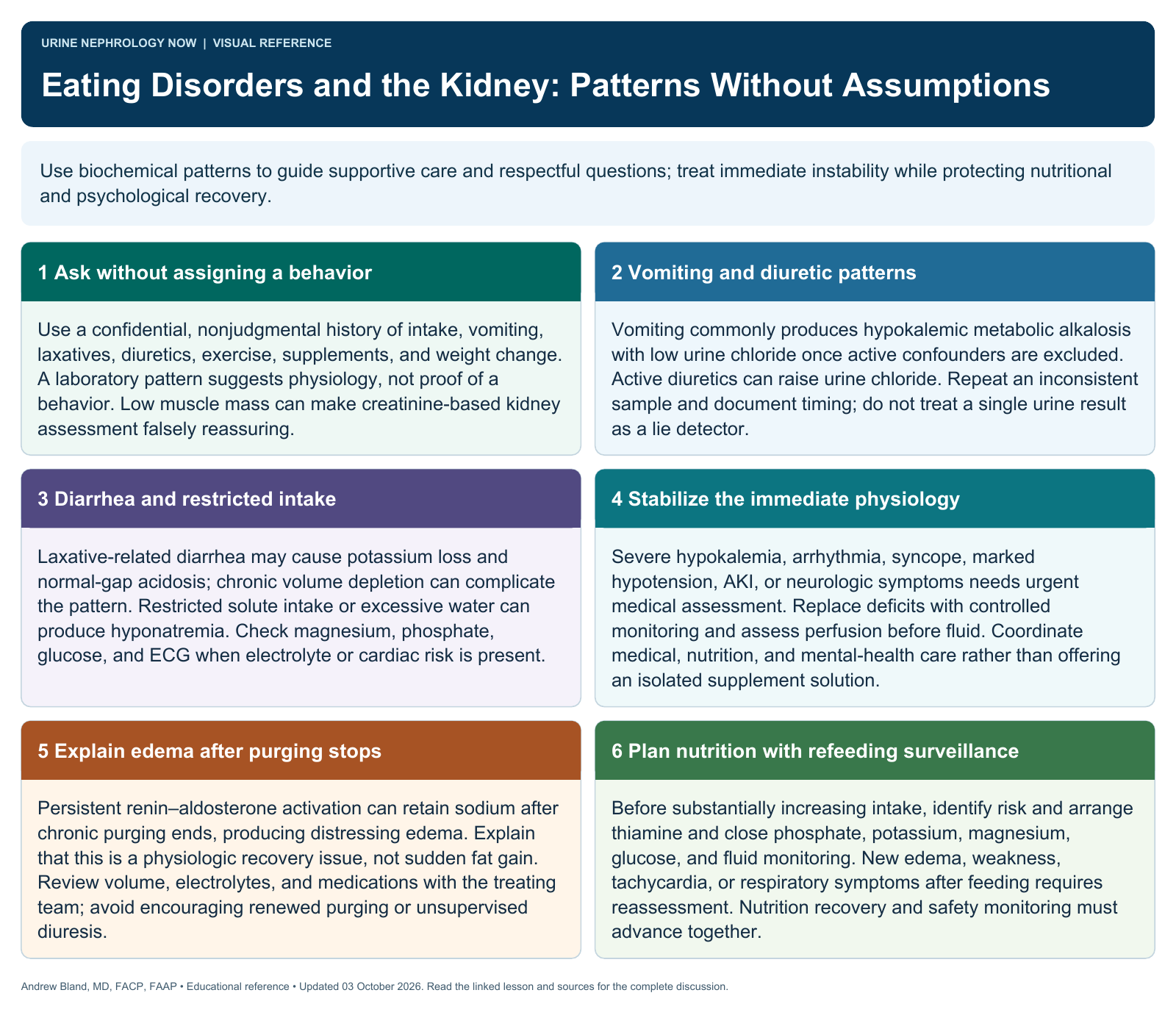
The specific purging method determines the acid-base and electrolyte pattern. Recognizing these signatures is essential for both diagnosis and management.
Self-Induced Vomiting
Most common purging method
- Metabolic alkalosis (loss of HCl from stomach)
- Hypokalemia (renal K wasting from alkalosis + aldosterone)
- Hypochloremia (direct loss of Cl in vomitus)
- Volume depletion → secondary hyperaldosteronism
- Urine Cl <20 mEq/L (chloride-responsive alkalosis)
- Elevated serum bicarbonate
- Hyponatremia (if polydipsia coexists)
Laxative Abuse
Second most common purging method
- Non-anion gap metabolic acidosis (GI loss of bicarbonate)
- Hypokalemia (GI loss of potassium in diarrhea)
- Volume depletion
- Urine Cl is variable
- Hypomagnesemia (GI losses)
- Hypernatremia if dehydration predominates
- Stool osmotic gap elevated with osmotic laxatives
Diuretic Abuse
Third purging method
- Metabolic alkalosis (contraction alkalosis + distal H+ secretion)
- Hypokalemia (renal K wasting)
- Volume depletion
- Urine Cl >20 mEq/L during active use (chloride-resistant pattern)
- Urine Cl <20 mEq/L after diuretic effect wears off
- Hyponatremia (thiazides), hypocalcemia (loops)
- Hypomagnesemia (both loop and thiazide)
📋 Summary: Electrolyte Signature by Purging Method
| Feature | Vomiting | Laxative Abuse | Diuretic Abuse |
|---|---|---|---|
| Acid-base | Metabolic alkalosis | Non-AG metabolic acidosis | Metabolic alkalosis |
| Potassium | Low | Low | Low |
| Chloride (serum) | Low | Variable | Low |
| Urine Cl | <20 mEq/L | Variable | >20 (active) / <20 (off) |
| Volume status | Depleted | Depleted | Depleted |
| Other clues | Dental erosion, parotid enlargement, Russell sign | Melanosis coli, stool osm gap | Urine diuretic screen positive |
🔬 Urine Chloride: The Key Diagnostic Clue
In the evaluation of metabolic alkalosis, the urine chloride is the single most useful test to distinguish etiologies and guide treatment. It is the nephrologist's secret weapon when an eating disorder is suspected.
💡 Why Urine Chloride, Not Urine Sodium?
In metabolic alkalosis, large amounts of bicarbonate are filtered. The kidney reabsorbs sodium with bicarbonate (as NaHCO3), so urine sodium can be misleadingly high even in volume depletion. Urine chloride is NOT affected by bicarbonaturia and accurately reflects the volume status.
"Chloride-Responsive"
Volume-depleted
Responds to NS resuscitation
Causes:
Vomiting / NG suction
Remote diuretic use
Post-hypercapnic
"Chloride-Resistant"
Does NOT respond to NS alone
Causes:
Active diuretic use
Bartter / Gitelman syndrome
Mineralocorticoid excess
Severe hypokalemia
💡 The Vomiting Patient: Step-by-Step Renal Physiology
- HCl is lost in vomitus → plasma HCO3 rises → metabolic alkalosis
- Volume depletion from fluid losses → activates RAAS
- Aldosterone rises → enhanced Na reabsorption in collecting duct in exchange for K+ and H+
- Renal K wasting → hypokalemia (from both alkalosis-driven intracellular shift AND aldosterone-driven renal loss)
- Cl depletion → kidney cannot reabsorb NaHCO3 without Cl → alkalosis is "maintained" by Cl deficit
- Paradoxical aciduria: Despite systemic alkalosis, severe hypokalemia can cause the kidney to secrete H+ instead of K+ → acidic urine in an alkalotic patient
🔬 Pseudo-Bartter Syndrome
Pseudo-Bartter syndrome refers to a clinical and biochemical picture that mimics Bartter syndrome but is caused by an identifiable external cause (most commonly chronic purging). It is one of the most important nephrology diagnoses to recognize in eating disorder patients.
📋 Features of Pseudo-Bartter Syndrome
| Feature | True Bartter Syndrome | Pseudo-Bartter (from Purging) |
|---|---|---|
| Pathophysiology | Genetic defect in loop of Henle transporters | Chronic volume depletion from purging → secondary hyperaldosteronism |
| Metabolic alkalosis | Yes | Yes |
| Hypokalemia | Yes | Yes |
| Elevated renin/aldosterone | Yes | Yes |
| Urine Cl | >20 mEq/L (renal Cl wasting) | <20 mEq/L (if from vomiting) or variable |
| Blood pressure | Normal to low | Normal to low |
| Age of onset | Childhood | Adolescence/young adulthood |
| Key distinguishing clue | Persistent renal Cl wasting | History, physical signs (Russell sign, dental erosion), urine Cl <20 |
⚠️ The Edema Rebound
When chronic purging stops, patients develop significant peripheral edema that can last 1-2 weeks. This occurs because:
- Chronic volume depletion has caused sustained RAAS activation
- When purging stops, the kidneys continue to avidly retain sodium and water
- This edema is frightening to the patient and often triggers relapse into purging
- Management: Reassurance that edema is temporary; spironolactone 25-50 mg can help; avoid diuretics (may worsen the cycle)
🔴 Bulimia Nervosa: Renal & Metabolic Complications
Bulimia nervosa is characterized by recurrent episodes of binge eating followed by compensatory behaviors (purging, fasting, excessive exercise). The renal complications stem primarily from purging behaviors and chronic volume depletion.
📋 Metabolic Complications of Bulimia
Electrolyte Disturbances
- Hypokalemia — most dangerous; risk of fatal arrhythmia (QT prolongation, U waves, ventricular tachycardia)
- Metabolic alkalosis — from HCl loss
- Hypomagnesemia — contributes to refractory hypokalemia
- Hypophosphatemia — especially during refeeding
- Hyponatremia — if excessive water intake accompanies purging
Renal Complications
- Prerenal AKI from chronic volume depletion
- Hypokalemic nephropathy — chronic tubulointerstitial injury from sustained hypokalemia
- Nephrocalcinosis — calcium phosphate deposition from chronic alkalosis
- Kidney stones — uric acid (dehydration, concentrated urine) and calcium oxalate
- Impaired urinary concentrating ability (NDI-like from chronic hypokalemia)
- CKD — from chronic tubulointerstitial disease if untreated for years
💡 Physical Exam Clues in Bulimia
- Russell sign: Calluses or scars on the dorsum of the hand/knuckles from inducing vomiting
- Dental erosion: Perimolysis — loss of enamel on lingual surfaces of teeth from repeated acid exposure
- Parotid gland enlargement: Bilateral, painless; gives "chipmunk" facies
- Elevated serum amylase: Salivary (not pancreatic) origin from parotid hypertrophy
- Normal or near-normal BMI: Unlike anorexia, bulimia patients are often normal weight
🔵 Anorexia Nervosa: Renal & Metabolic Complications
Anorexia nervosa is characterized by severe caloric restriction, distorted body image, and fear of weight gain. BMI is typically <17.5 kg/m² (or <85% of expected weight). The renal complications result from malnutrition, volume depletion, and the dangers of refeeding.
📋 Renal Complications of Anorexia
Acute Complications
- Prerenal AKI — dehydration, reduced cardiac output, muscle wasting reducing creatinine production (may mask true GFR decline)
- Low serum creatinine — reflects muscle wasting, NOT good kidney function; eGFR may be falsely reassuring
- Elevated BUN:Cr ratio — prerenal pattern
- Nephrogenic diabetes insipidus — impaired concentrating ability from chronic malnutrition/hypokalemia
Chronic Complications
- Chronic tubulointerstitial disease from prolonged hypokalemia
- Nephrocalcinosis
- Kidney stones (concentrated urine, low citrate)
- Reduced GFR — reported in up to 10% of chronic AN patients
- Osteoporosis → hypercalciuria → stone risk
💡 The Creatinine Trap in Anorexia
Serum creatinine in severe anorexia may be deceptively low (0.3-0.5 mg/dL) due to profound muscle wasting. A creatinine of 0.8 mg/dL in a severely cachectic patient may represent significant kidney injury. Cystatin C-based eGFR may provide a more accurate assessment of true GFR in this population.
⚠️ Refeeding Syndrome
Refeeding syndrome is a potentially fatal complication that occurs when nutrition is restarted in severely malnourished patients. It is characterized by dangerous shifts in electrolytes driven by insulin release and the resumption of anabolic metabolism.
📋 Pathophysiology of Refeeding Syndrome
- Starvation state: Body depletes intracellular stores of phosphorus, potassium, and magnesium, but serum levels may appear normal (total body depletion masked by low intake and reduced cellular uptake)
- Carbohydrate reintroduction: Insulin secretion surges
- Insulin drives cellular uptake of phosphorus, potassium, magnesium, and glucose
- Serum levels plummet: Severe hypophosphatemia, hypokalemia, hypomagnesemia
- Consequences: Cardiac arrhythmia, respiratory failure (diaphragmatic weakness from hypophosphatemia), rhabdomyolysis, seizures, death
⚠️ Risk Factors for Refeeding Syndrome
- BMI <16 kg/m²
- Unintentional weight loss >15% in 3-6 months
- Little or no nutritional intake for >10 days
- Low baseline phosphorus, potassium, or magnesium before refeeding
- History of alcohol use disorder, chemotherapy, or chronic diuretic/laxative use
💊 Prevention & Management of Refeeding Syndrome
| Step | Action |
|---|---|
| 1. Check electrolytes | Phosphorus, potassium, magnesium, calcium, glucose BEFORE starting nutrition |
| 2. Replete FIRST | Correct phosphorus (>3.0), potassium (>4.0), and magnesium (>2.0) BEFORE refeeding |
| 3. Start low, go slow | Begin at 10-20 kcal/kg/day (or 50-75% of estimated needs); advance by 200 kcal/day every 1-2 days |
| 4. Monitor aggressively | Check phosphorus, K, Mg, glucose every 6-12 hours for first 72 hours, then daily x 5-7 days |
| 5. Thiamine | Give thiamine 200-300 mg IV before starting carbohydrates (prevents Wernicke encephalopathy) |
| 6. Fluid management | Restrict IV fluids; avoid excessive sodium loads that can precipitate fluid overload in a patient with impaired cardiac function |
⚠️ Phosphorus Is the Sentinel Electrolyte
Hypophosphatemia is the hallmark of refeeding syndrome and the earliest laboratory marker. A falling phosphorus within 24-72 hours of starting nutrition should prompt immediate slowing of caloric delivery and aggressive phosphorus repletion. Severe hypophosphatemia (<1.0 mg/dL) can cause respiratory failure from diaphragmatic weakness, hemolytic anemia, and cardiac arrest.
🏥 Chronic Renal Complications of Eating Disorders
📋 Long-Term Kidney Effects
Nephrocalcinosis
- Calcium phosphate deposition in renal medulla
- Driven by chronic alkalosis (increases urinary calcium excretion) and hypokalemia
- Visible on renal ultrasound or CT
- Can cause chronic tubulointerstitial nephritis
- May be partially reversible if eating disorder is treated early
Kidney Stones
- Uric acid stones: Concentrated, acidic urine (especially in laxative abuse with low urine volume)
- Calcium oxalate stones: Low urine volume, low citrate
- Calcium phosphate stones: Chronic alkalosis, hypercalciuria
- Risk amplified by chronic dehydration and low urine output
Chronic Tubulointerstitial Disease
- Hypokalemic nephropathy: Chronic hypokalemia causes vacuolization of proximal tubular cells, interstitial fibrosis, and tubular atrophy
- Impaired urinary concentrating ability (resembles nephrogenic DI)
- Polyuria and polydipsia
- May progress to CKD stage 3-4 with years of untreated disease
- Partially reversible with sustained potassium correction
📋 Screening Questions for the Nephrologist
When the electrolyte pattern suggests an eating disorder, sensitive and non-judgmental screening is essential. Patients rarely volunteer this history.
💬 The SCOFF Questionnaire
A validated 5-question screen (2 or more "yes" answers = positive screen):
- Sick: Do you make yourself sick because you feel uncomfortably full?
- Control: Do you worry you have lost control over how much you eat?
- One stone: Have you recently lost more than one stone (14 lb / 6.4 kg) in a 3-month period?
- Fat: Do you believe yourself to be fat when others say you are too thin?
- Food: Would you say that food dominates your life?
📋 Practical Approach for the Nephrologist
When the lab pattern is suspicious, consider asking:
- "I'm seeing a pattern in your blood work that I sometimes see in patients who are having trouble with eating or using methods to control their weight. Can we talk about that?"
- "Have you been using any over-the-counter products like laxatives, diuretics, or diet pills?"
- "Have you been having any vomiting or nausea?"
- "How would you describe your relationship with food and your body?"
🔬 Confirmatory Laboratory Workup
| Test | Purpose |
|---|---|
| BMP (Na, K, Cl, HCO3, BUN, Cr, Glucose) | Identify acid-base pattern, hypokalemia, AKI |
| Magnesium, phosphorus, calcium | Often co-depleted; Mg needed to correct K |
| Urine chloride | Key: <20 mEq/L = vomiting; >20 = diuretic use or Bartter |
| Urine potassium (spot K/Cr ratio or TTKG) | Assess renal vs extrarenal K loss |
| Urine diuretic screen | Detects loop diuretics, thiazides, acetazolamide |
| Urine laxative screen (if available) | Detects bisacodyl, senna, phenolphthalein |
| Amylase (with isoenzymes) | Elevated salivary amylase suggests purging |
| ECG | QT prolongation, U waves, ST changes from hypokalemia |
💊 Treatment Approach for the Nephrologist
📋 Acute Management: Electrolyte Replacement
| Electrolyte | Replacement Strategy | Monitoring |
|---|---|---|
| Potassium |
Oral KCl preferred (40-80 mEq/day in divided doses). IV KCl for K <3.0 or symptomatic (max 10-20 mEq/hr via central line with telemetry). Correct magnesium first — hypokalemia is refractory until Mg is repleted. |
Recheck q4-6h until stable. ECG monitoring if K <2.5. |
| Magnesium |
MgSO4 1-2g IV over 1-2 hours (for severe depletion). Oral Mg oxide 400-800 mg/day for maintenance. |
Recheck 24h after replacement. Body stores replete slowly; plan for 3-5 days of supplementation. |
| Phosphorus |
IV sodium/potassium phosphate 15-30 mmol over 6h (for P <1.5 or symptomatic). Oral Neutra-Phos 1-2 packets TID for mild depletion. |
Recheck q6h during refeeding. Watch for hypocalcemia from phosphate infusion. |
💧 Volume Restoration
- Isotonic saline (0.9% NaCl) is the initial fluid of choice for volume depletion
- The chloride in NS corrects the chloride deficit, which is essential for resolving the metabolic alkalosis
- Avoid aggressive fluid resuscitation in malnourished patients — compromised cardiac function may lead to pulmonary edema
- Target correction: 1-2 L over 12-24 hours, guided by clinical assessment
- Acetazolamide (250-500 mg IV/PO) can be used for severe alkalosis (pH >7.55 or HCO3 >40) to promote renal bicarbonate excretion, but only after volume and chloride are repleted
🧠 Psychiatric Referral & Multidisciplinary Care
- Psychiatric referral is essential — electrolyte correction without addressing the underlying disorder leads to recurrence
- Cognitive behavioral therapy (CBT) is first-line for bulimia nervosa
- Family-based therapy (FBT/Maudsley method) is first-line for adolescent anorexia
- SSRIs (fluoxetine) have evidence for bulimia (reduces binge/purge frequency)
- Coordinate with dietitian for structured meal planning and refeeding protocols
- Criteria for inpatient psychiatric admission: BMI <15, K <2.5, active suicidality, hemodynamic instability, refeeding syndrome
💡 The Nephrologist's Unique Role
The nephrologist may be the first provider to identify the eating disorder through the electrolyte pattern. Our role includes:
- Recognition: Identify the electrolyte signature and initiate the conversation
- Stabilization: Correct life-threatening electrolyte abnormalities and manage AKI
- Refeeding safety: Guide electrolyte monitoring during nutritional rehabilitation
- Long-term monitoring: Follow renal function and watch for chronic complications
- Referral: Connect the patient with psychiatry, psychology, and nutrition services
🎯 Key Learning Points
- Self-induced vomiting causes metabolic alkalosis, hypokalemia, and urine Cl <20 mEq/L.
- Laxative abuse causes non-anion gap metabolic acidosis (not alkalosis) with hypokalemia.
- Diuretic abuse causes metabolic alkalosis with urine Cl >20 mEq/L during active use.
- Urine chloride is the most useful single test to distinguish causes of metabolic alkalosis and to differentiate vomiting from diuretic abuse.
- Pseudo-Bartter syndrome from chronic purging mimics Bartter syndrome with hypokalemia, alkalosis, and secondary hyperaldosteronism.
- In anorexia, serum creatinine is deceptively low due to muscle wasting — do not be falsely reassured by a "normal" creatinine.
- Refeeding syndrome is caused by insulin-driven cellular uptake of phosphorus, potassium, and magnesium. Phosphorus is the sentinel marker.
- "Start low, go slow" with caloric reintroduction; replete electrolytes BEFORE refeeding.
- Chronic eating disorders can cause hypokalemic nephropathy, nephrocalcinosis, kidney stones, and CKD.
- Correct magnesium first — hypokalemia is refractory until magnesium is repleted.
- The SCOFF questionnaire is a validated screening tool for eating disorders.
- Psychiatric referral is essential — electrolyte correction alone does not treat the underlying disease.
📚 References
- Bouquegneau A, Dubois BE, Krzesinski JM, Delanaye P. Anorexia nervosa and the kidney. Am J Kidney Dis. 2012;60(2):299-307. PubMed: 22609034
- Stheneur C, Bergeron S, Lapeyraque AL. Renal complications in anorexia nervosa. Eat Weight Disord. 2014;19(4):455-460. PubMed: 24997544
- Bahia A, Mascolo M, Gaudiani JL, Mehler PS. PseudoBartter syndrome in eating disorders. Int J Eat Disord. 2012;45(1):150-153. PubMed: 21344464
- Mehler PS, Walsh K. Electrolyte and acid-base abnormalities associated with purging behaviors. Int J Eat Disord. 2016;49(3):311-318. PubMed: 26876281
- Friedli N, Stanga Z, Sobotka L, et al. Revisiting the refeeding syndrome: results of a systematic review. Nutrition. 2017;35:151-160. PubMed: 28087222
- Mascolo M, Trent S, Colwell C, Mehler PS. What the emergency department needs to know when caring for your patients with eating disorders. Int J Eat Disord. 2012;45(8):977-981. PubMed: 22707235
- National Institute for Health and Care Excellence (NICE). Eating disorders: recognition and treatment (NG69). 2017; updated 2020.
- Gaudiani JL, Sabel AL, Mascolo M, Mehler PS. Severe anorexia nervosa: outcomes from a medical stabilization unit. Int J Eat Disord. 2012;45(1):85-92. PubMed: 22170021
- Morgan JF, Reid F, Lacey JH. The SCOFF questionnaire: assessment of a new screening tool for eating disorders. BMJ. 1999;319(7223):1467-1468. PubMed: 10582927
- Palmer BF, Clegg DJ. Electrolyte and acid-base disturbances in patients with diabetes mellitus. N Engl J Med. 2015;373(6):548-559. PubMed: 26244308