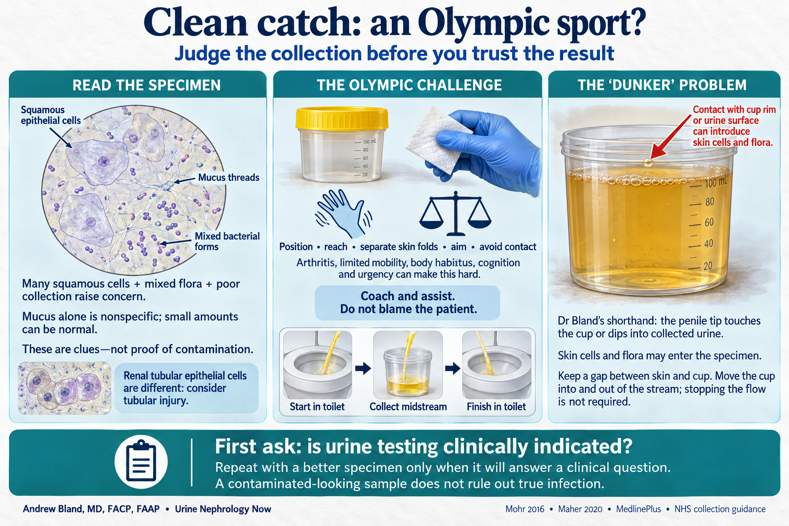
Squamous cells, mucus and mixed flora are collection clues. Read them with the collection method and clinical syndrome. The microscopy drawings are teaching schematics, not diagnostic photomicrographs.
Collection, microscopy and clinical context
Read the collection clues together
| Finding | What it tells you—and what it cannot prove |
|---|---|
| Many squamous epithelial cells | Large, flat cells from distal urethral or external genital surfaces can flag carryover during collection. They reduce confidence in interpreting a UA, but do not reliably establish culture contamination alone. Use the laboratory’s reporting units and reference range; there is no universal yes/no cutoff. |
| Mucus threads | Small amounts can occur normally. Abundant mucus may accompany genital secretions or urinary inflammation. Together with many squamous cells, mixed organisms and a difficult collection it adds context; mucus alone diagnoses neither contamination nor UTI. |
| Mixed bacterial forms or mixed growth | A mixture can suggest external flora, especially after pad, bag or skin contact. True polymicrobial infection can occur in selected settings, particularly with instrumentation or catheters; interpret the clinical setting and culture report. |
| White cells and leukocyte esterase | These indicate inflammatory cells in the sample, not necessarily bacterial infection in the bladder. Genital carryover and asymptomatic bacteriuria can also produce an abnormal UA. Neither pyuria nor bacteria alone establishes symptomatic UTI. |
| Renal tubular epithelial cells | Do not put every epithelial cell in the contamination bucket. Renal tubular epithelial cells, especially with granular casts, support a tubular injury process; they are different from large squamous cells. |
Why a clean catch can feel like an Olympic sport
“Clean catch is an Olympic sport” is a memorable way to describe the task: position safely, reach, separate skin folds, clean, aim, collect midstream and keep the cup away from the body—all while needing to urinate. Arthritis, limited mobility, body habitus, visual difficulty, urgency and cognitive impairment can make that coordination unrealistic. Ask what the patient can actually do, give clear instructions, provide a suitable setup and offer assistance. A difficult collection is a practical care problem, not a patient failure.
The “dunker” problem—and a practical fix
“Dunkers” is Dr Bland’s informal teaching term for collection in which the penile tip touches the cup or dips into already collected urine, allowing external cells and flora into the specimen. Urgency, dribbling, obstructive symptoms or difficulty positioning the cup may contribute; BPH does not make this inevitable. Keep a gap between skin and container. Midstream collection does not require stopping and restarting the flow: begin into the toilet, move the cup into the stream, then remove it and finish into the toilet.
Coach the collection
Wash hands and prepare the supplied container. Follow local cleansing instructions; separate the labia or retract the foreskin as appropriate. Avoid touching the inside of the cup or lid. Collect the middle portion without contacting the skin, cap it and return it promptly; use the laboratory’s storage instructions if transport is delayed.
Repeat only when there is a clinical question
First decide whether urine testing will answer a clinical question. If a specimen is needed and collection quality is poor, repeat with coached or assisted midstream collection; consider a trained clinician’s in-and-out catheter specimen when a reliable sample is clinically necessary and clean catch is not feasible. Catheterization is not a routine response to incidental pyuria or bacteriuria. A contaminated-looking specimen can coexist with true infection, and suspected sepsis requires prompt assessment and treatment while the source is evaluated.
Sources
Day 3 · Case 7 teaching before Q4 · Urinalysis interpretation lecture