Visual summary
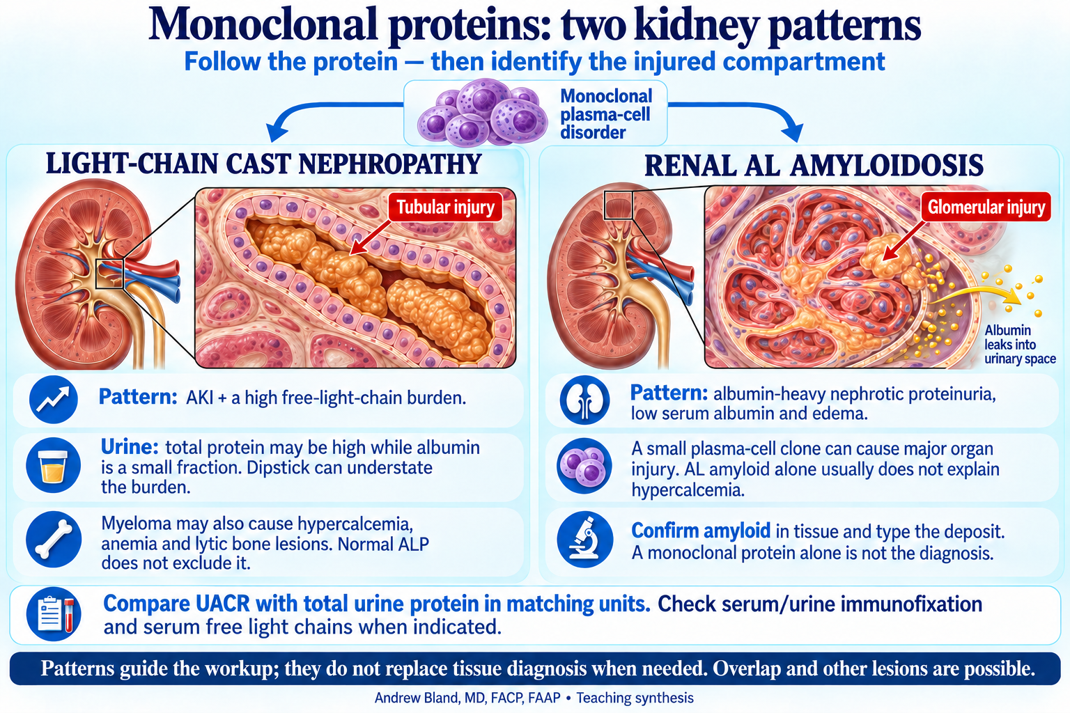
Tubular light-chain cast injury versus glomerular AL amyloid deposition.
Teaching text
Follow the protein into the kidney
Monoclonal immunoglobulin disease can injure different kidney compartments. Cast nephropathy mainly affects tubules; AL amyloid often affects glomeruli and can also involve vessels and interstitium. These are two important patterns, not the entire monoclonal kidney differential.
Tubular cast pattern
A large total urine protein burden with a much smaller albumin component suggests overflow light chains or another nonalbumin source. Dipsticks mainly detect albumin and can understate this burden. Markedly abnormal serum free light chains and AKI raise concern for cast nephropathy; the pattern is not histologic proof.
Glomerular amyloid pattern
Albumin-dominant nephrotic proteinuria, low serum albumin and edema suggest glomerular injury. In AL amyloidosis the clone can be small. Confirm amyloid in tissue and type the deposit; a monoclonal protein alone does not establish AL. Kidney biopsy is one option when clinically indicated, not a requirement for every patient if another tissue establishes the diagnosis.
Link the findings without conflating them
Myeloma can cause hypercalcemia from bone resorption, anemia, lytic lesions and multiple kidney injuries. AL amyloid alone does not usually explain hypercalcemia or lytic lesions. A patient can have overlapping lesions, and other monoclonal diseases such as light-chain deposition disease remain possible.
Tests and escalation
Use serum and urine immunofixation, electrophoresis and serum free light chains when indicated. CKD alters absolute light-chain levels and assay interpretation. Match UACR and total protein using the same units; this is a clue, not a substitute for urine protein electrophoresis or biopsy. Suspected myeloma-related AKI needs prompt hematology and nephrology coordination.
Continue learning
Read the connected teaching · Summary-day capstone · Bring the lecture to the bedside